Bovine larynx: Actinobacillosis.Detailed information
Animal: Bovine, 11 years, Norwegian red cattle, female
Organ: Laryngeal mass
History: The cow developed intermittent, moderate respiratory distress over a period of approximately 2 weeks. Otherwise normal clinical signs.
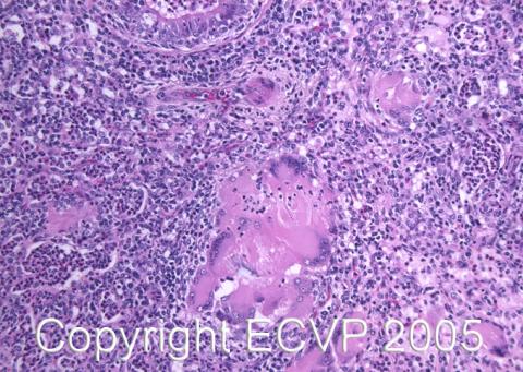
Autopsy findings: In the ventral larynx, distal to the epiglottis, a mass measuring 5 x 3 x 2 cm was located. It had a bumpy, non-ulcerated surface and seemed to grow beneath the mucosa. The cut surface was lobulated, with areas of yellow-brown, caseous tissue surrounded by a firmer, greyish to white tissue. When pressure was applied to the sample, tiny, yellow granules appeared on the surface. Histological examination of the tissue revealed multiple, purple colonies of bacteria surrounded by a mixture of neutrophils, macrophages and multinucleated giant cells in a collagen rich stroma. The purple colonies had club shaped structures radiating out on the surface.
Diagnosis: Larynx, pyogranulomatous inflammation with intralesional club colonies consistent with Actinobacillus lignieresii. The agent was not isolated upon cultivation.Necropsy performed by: Gjermund Gunnes Photo by: Gjermund Gunnes