Dog, Boxer: Brain, lateral ventricles: choroid plexus carcinoma with neuronal atrophy and moderate internal hydrocephaly
History: Dog, Boxer, female, 8 years
Diagnosis: Brain, lateral ventricles: choroid plexus carcinoma with neuronal atrophy and moderate internal hydrocephaly
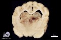
Description: Almost centrally and protruding dorsally from the midbrain into the lateral ventricles is a partially invasive mass measuring up to 3.0 x 2.6 x 3.7 cm. The cut face is beige in color with multifocal to coalescing, small, red-brown areas (hemorrhages) and a multifocally cystic appearance with cysts of up to 0.4 cm in diameter filled with a clear fluid. The cerebral cortex is narrowed (neuronal atrophy) and both lateral ventricles are moderately dilated.
Comments: This bitch was euthanized due to a major change in her behavior. She was atactic, had an urge to move forward, became aggressive and tried to bite people. Histologically, the mass was composed of a fibrovascular stroma covered by an epithelium consisting of cuboidal to columnar cells. Increased numbers of mitoses and invasive growth were present and some cells formed solid sheets while losing their papillary pattern.
This rare neoplasm most commonly occurs in male dogs with no predilection to a specific breed, but is occasionally also seen in horse and cattle. The most common localization is the fourth ventricle, but the neoplasm is also found in the third or the lateral ventricles.
Hydrocephaly is a common consequence due to either obstruction of the ventricular system and/or to an overproduction of liquor.
Picture and authored by: Hannah Pischon, Department of Veterinary Pathology, Freie Universität Berlin, Germany