Animal: Dog, male, 13 years
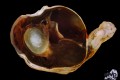
Diagnosis: Eye (right): Uveal melanoma with invasion beyond the sclera and into the optic nerve, retinal detachment, hyphema and cataract
Description: More than half of the globe is replaced by a brown to black mass enclosing the lens, invading or breaking through the sclera multifocally and spreading into the optic nerve. The retina is detached and multifocal hemorrhage especially into the anterior chamber (hyphema) is present.
Comments: The dog was presented clinically with severe keratitis and hyphema impeding a complete examination of the inner eye structures. The owner reported a reddening of the affected eye and anti-inflammatory treatment without success. The globe was exenterated. While primary ocular melanocytic tumors are common in dogs and cats, they are rare in horses, and almost nonexistent in other domestic animals. In dogs most of the uveal melanocytic tumors are benign melanocytomas mostly originating from the anterior uvea. Definite differentiation of benign melanocytomas from malignant melanomas is somewhat controversial as local invasion into and through the sclera is recorded for the benign variant as well. A preponderance of epitheloid, heavily pigmented, plump melanocytes in contrast to lightly pigmented spindle cells is indicative of benign melanocytoma. Nuclear anaplasia including anisokaryosis, karyomegaly, folded nuclei, and large nucleoli as well as mitotic index greater than four mitotic figures per 10 high power fields are characteristics of malignant melanoma.
Authored and photo by Olivia Kershaw, Department of Veterinary Pathology, Freie Universität Berlin, Germany