Animal: Dog, 5 years, female.
Diagnosis: Thymus, heart and large vessels: thymic carcinoma.
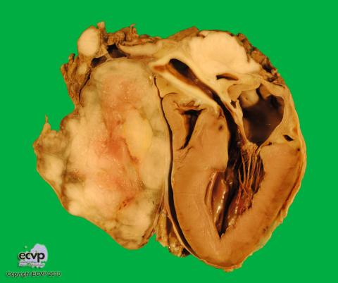
Description: The pre- and supracardial mass measured up to 12.9 cm in diameter and was multilobulated, grey to beige and with a greasy consistence. The aortic and pulmonary root vessels were completely encircled by the neoplasm. Additionally, tumor cells infiltrated the vessel walls as well as the adjacent adipose tissue, pericard and epicard.
Comments: Histological examination identified the neoplasm as a malignant proliferation of thymic epithelial cells with a marked, lymphocytic infiltration which is considered to be due to IL-2 secretion of the epithelial tumour cells. Thymic carcinomas develop in middle-aged dogs but are less frequent than thymic malignant lymphomas. Only occasionally do such tumours spread through distant metastases but interference with cardiac function is more commonly observed. In the case presented here, only the heart was submitted for pathological investigation and no further information regarding metastases was available.
Picture by: Marie-Charlotte von Deetzen, Department of Veterinary Pathology, Freie Universität Berlin, Germany
Author: Marie-Charlotte von Deetzen, Department of Veterinary Pathology, Freie Universität Berlin, Germany