Diagnosis: dog, third phalanx, subungual epithelial inclusion cyst
Animal: Tibetan terrier, 10 years, male
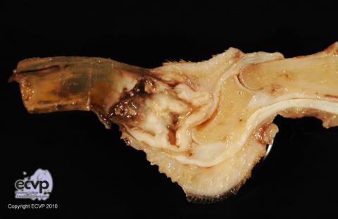
Organ: Third phalanx
History: A distal extremity of the dog was submitted without case history.
Macroscopical findings: The dactyl was enlarged and laterally flattened. Within the third phalanx the bone was replaced by a cystic cavity.
Histopathological findings: Within the third phalanx an expansive cystic cavity was found, which replaced the bony tissue. The cystic cavity was lined by a well differentiated keratinizing multilayered epithelium with pyogranulomatous inflammation in the adjacent tissue.
Diagnosis: dog, third phalanx, subungual epithelial inclusion cyst
Investigation and photo by: Vanessa Herder, Andreas Beineke, Department of Pathology, University of Veterinary Medicine Hannover Foundation, Germany