Dog eye: Ocular medulloepithelioma
Animal: Dog, 4-year-old Neapolitan mastiff, female.
Organ: Eye.
History: The dog presented with acute glaucoma of the left eye and swollen hock joints. The dog was depressed and had poor conscious proprioception of both hind limbs. Examination of the left eye revealed peripheral corneal vascularization and severe corneal oedema. The depression became more pronounced during the following week. CNS signs progressed rapidly and the dog got seizures and went into a coma and was subsequently euthanazied.
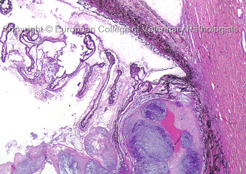
Description: The ciliary body contained an unpigmented tumour mass. The tumour tissue was greyish-white, gelatinous and semitransparent with scattered opaque white foci. The neoplastic tissue protruded into the posterior chamber and vitreous, partly surrounding the lens. In the brain, the cerebral ventricles contained soft, grayish, lucent tissue. A wedge-shaped, grey and soft area was observed in one kidney.
Histology showed a multinodular, poorly differentiated tumor arising from the ciliary body. The neoplastic tissue was composed of small islets and cords of nonpigmented, pleomorphic cells. Multifocally chondroid differentiation was observed. Tumour cells were surrounded by abundant, amorpous ground substance. Neoplastic cells with similar morphology were present in the brain ventricles and the grey area in the kidney.
Diagnosis: Ocular medulloepithelioma with distant metastasis
Necropsy performed by: Dr. Mona Aleksandersen
Photo by: Dr. Mona Aleksandersen.