Diagnosis: Tiger, lens, cataract, mature to hypermature, posterior
Animal: Panthera tigris, 19 years, male.
Organ: lens
History: The captive tiger showed bilateral lenticular opacity for several years. One year prior death the animal loosed weight and displayed a poor general condition with progressive depression, anorexia and recumbency. Due to a poor prognosis the tiger was euthanized and submitted to necropsy.
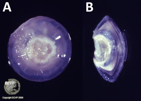
Macroscopical findings: Both lenses displayed degeneration and white discoloration with a circular white wrinkling on the posterior pole (A). Cross section revealed a lenticular degeneration, which extended from the posterior pole to the equator of the lens (B).
Histopathological findings: Both lenses showed a severe degeneration and liquefaction of lens fibers with sclerosis and an eccentric nucleus. Dystrophic mineralization as well as Morgagnian globules and a slight posterior migration of lens epithelium were visible.
Comment: Further necropsy findings included multiple, mediastinal and metastizing mesotheliomas with serous pleural effusion. In addition, chronic lymphocytic interstitial nephritis, bilateral parathyroidal hyperplasia as well as metastatic mineralizations were observed.
Diagnosis: lens, cataract, mature to hypermature, posterior
Necropsy performed by: Vanessa Herder, Department of Pathology, University of Veterinary Medicine Hannover, Germany
Photo by: Vanessa Herder, Andreas Beineke