History: Beagle dog, female, 5 years
Diagnosis: Heart: Moderate, chronic, diffuse, right ventricular hypertrophy with intraluminal nematodes consistent with Dirofilaria immitis
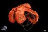
Description: The right ventricle contained four approximately 27 cm long, slender nematodes, embedded in a postmortem clot. The relative heart weight was within normal limits but the right ventricular wall was mildly thickened.
Comments: The bitch died due to complications after ovariohysterectomy, the adult heartworms were an incidental finding. Clinical signs attributable to the nematodes were absent. In gross and histological post mortem examination, besides the myocardial hypertrophy, sequelae commonly seen in cases of dirofilariasis including vasculitis with consecutive pulmonary hypertension, pulmonary fibrosis, congestive heart failure, vena cava syndrome or edema were absent.
Dirofilaria immitis is endemic in most mediterranean and southeastern european countries. Generally, it is a cosmopolitan infection present in most countries with temperate, semitropical or tropical climates as it is transmitted by over 70 different species of mosquitoes.
In domestic animals Dirofilaria frequently causes disease in dogs and less frequently in cats and ferrets. It can also cause patent infection in numerous wild mammalian species, including wolves, coyotes, raccoons, foxes, seals, sea lions and even more species can be infected non-patently, including humans and horses. Additionally, dead larvae may cause pulmonary granulomatous vasculitis, however usually without other relevant consequences in dogs.
Picture by: Stefanie Binder, Department of Veterinary Pathology, Freie Universität Berlin, Germany
Authored by: Hannah Pischon, Department of Veterinary Pathology, Freie Universität Berlin, Germany