Animal: karakul sheep (Ovis aries), 3 years, female
Organ: Esophagus
History: The sheep collapsed with no prior clinical signs. After subsequent, separate housing, the general condition deteriorated so that the animal had to be euthanized.
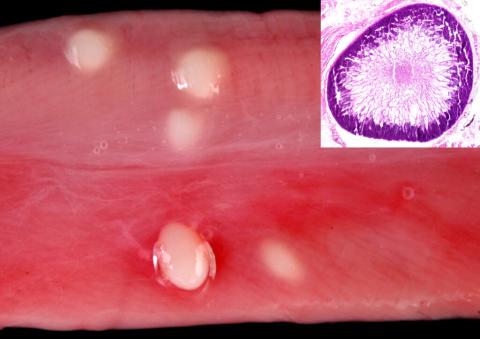
Autopsy findings: The animal was in a cachectic nutritional state and showed an intestinal endoparasitosis with various nematode species. Three tapeworm fins (metacestode of Taenia hydatigena) were found in the abdominal cavity. Multiple creamy white to pale yellow parasitic structures up to 0,5 cm in size were identified in the esophageal serosa.
Histopathological findings: The cysts were surrounded by a thin eosinophilic capsule. Numerous septa extend from the capsule to the center of the cysts. The cysts also contain numerous basophilic bradyzoites.
Diagnosis: Esophagus: multifocal, nonreactive sarcosporidial cysts (Etiology: Sarcocystis gigantea)
Contributors: Christiane Helm (Institute of Veterinary Pathology, Faculty of Veterinary Medicine, Leipzig University, Germany), Ronald Schmäschke (Institute of Veterinary Parasitology, Faculty of Veterinary Medicine, Leipzig University, Germany), Florian Hansmann (Institute of Veterinary Pathology, Faculty of Veterinary Medicine, Leipzig University, Germany)
Pictures: Christiane Helm (Institute of Veterinary Pathology, Faculty of Veterinary Medicine, Leipzig University, Germany)