History: Cat, British short hair, 15 years, female
Diagnosis: Vertebral column with spinal cord (L6 – S3): Meningioma with compression of the spinal cord, partial displacement and hyperosteosis of adjacent vertebral bodies
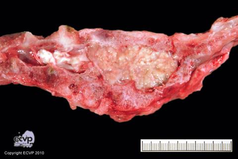
Description: Paramedian sagittal section of the caudal vertebral column (L6 – S3): The spinal cord and vertebrae are compressed and partly replaced by an expansile growing, soft, brown to whitish mass in the spinal canal with finely lobular appearance.
Comments: The cat presented with neurological deficits affecting both hind legs and progressive generalized movement disorder. Necropsy revealed an expansile, soft, whitish, 3.5 x 2.5 x 2.0 cm mass expanding the spinal canal. The mass extended from L6 to S3 and compressed or replaced the spinal cord and vertebral bone. Histologically, spindle shaped to polygonal tumour cells, arranged in whorls with multifocal, centrally located “psammoma bodies“ led to the diagnosis of a meningioma. Meningiomas are frequently seen in older cats and mainly arise from the meninges within the cranium but are rarely seen along the spinal cord.
Picture by: Anja Meyer, Department of Veterinary Pathology, Freie Universitaet Berlin, Germany
Author: Anja Meyer, Department of Veterinary Pathology, Freie Universitaet Berlin, Germany