Animal: Dog, Scottish terrier, 3 years, female
Diagnosis: Cross section of the heart, left ventricle and septum: Lymphoma, multifocal to coalescing
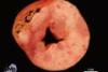
Description: The left ventricle and the septum were thickened with a multifocal to coalescing, white to beige-coloured masses mostly replacing the myocardium with a maximum extension of 3.2 x 2.7 x 1.1 cm. The borders of the masses were irregular and poorly circumscribed. Additionally, multifocal petechial hemorrhages were present.
Comments: The Scottish terrier was clinically presented with sudden onset of severe dyspnoea. After intubation, she started bleeding from the endotracheal tube. She died spontaneously. Necropsy revealed multifocal white to beige-coloured masses in various organs including the heart, lung, diaphragm and liver. Additionally, severe, acute, multifocal to coalescing haemorrhages in the lung and moderate, acute, multifocal haemorrhages in the meninx were present. Histopathological examination revealed an infiltration of the myocardium with neoplastic round cells that resembled lymphoblasts. Similar infiltrations were present in the lung and associated with multifocal, acute haemorrhages. Additionally, similar neoplastic round cells infiltrated the liver, diaphragm, kidneys and urinary bladder. Lymph nodes were not enlarged and representative sections of lymph nodes lacked tumor cells microscopically. Unexpectedly from the pathological findings, there were no pathological signs of a chronic cardiac insufficiency.
Lymphomas represent the most common haematopoietic neoplasm in dogs. The mean age of occurrence is between 5 and 9 years; but dogs younger than 6 months may also be affected. Immunohistochemical phenotyping can classify most lymphomas as of T or B cell origin. The updated Kiel system is a common classification scheme; however different classifications, such as the human WHO classification, are currently investigated.
In most cases multiple organs are involved and usually lymphatic tissue such as (superficial) lymph nodes, spleen and bone marrow are involved. Besides those multicentric lymphomas, other forms are occasionally present: alimentary, mediastinal, cutaneous and extranodal forms. However, lymphoma may be observed in almost each tissue of the body. Clinical signs are highly variable, depending on the extent and location and therefore may sometimes be vague. In 10 to 35 % of the cases, a hypercalcemia of malignancy may be observed as a result of a paraneoplastic syndrome, i.e., production of parathyroid hormone-related peptide. In dogs a multifactorial pathogenesis is suspected. An underlying retroviral infection as cause of lymphoma in cats and cattle is currently thought to not occur in dogs.
Picture by Stefanie Binder, Department of Veterinary Pathology, Freie Universitaet Berlin, Germany
Author: Christof Bertram, Department of Veterinary Pathology, Freie Universitaet Berlin, Germany