Animal: Dog, intact male, “Fila de S. Miguel” breed, 8 years old.
An accidentally found nodule was removed from the left pectoral region, at the time of sedation for amputation of thesupplementary fingers. Dimensions 1.9 x 1.2 x 1.2 cm. A mid-section was subject to decalcification prior to processing.
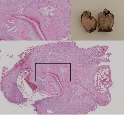
The macro photograph was taken from reserved material after the nodule had been analysed. A slice is missing parallel to theremaining fragments and corresponding to the processed material.
The fragment under analysis consisted of plates of well differentiated bone tissue, which surround a structure that fullyreproduces a nail bed in which keratin is formed from well differentiated squamous epithelium. One of the bone plates servesas a support to the epithelium like the same structure present in the digits. The nail is fully surrounded by skin folds in whichnumerous tubular structures are lodged with the morphology of the eccrine glands of the footpads.
Diagnosis was of hamartoma that developed in the pectoral skin, possibly during embryonal development, which fullyreproduces the structure of a digit with a nail. The fact that the nodule stayed unnoticed for so long possibly has to do with the fact that no rejection of a self-structure was expected and the small volume in a under the body region was not regularly palpated.
Hamartoma is a benign (not cancer) growth made up of an abnormal mixture of cells and tissues normally found in the area of the body where the growth occurs (National Cancer Institute – https://www.cancer.gov/publications/dictionaries/cancer-terms/def/hamartoma)
Authorship (text and photos) – Maria C. Peleteiro
VetPat, Rua dos Soeiros, 307ª, 1500-580 Lisboa, Portugal
mcpeleteiro@gmail.com