Animal: Baboon, > 20 years, female
Diagnosis: Large intestine: typhlocolitis, severe, acute, hemorrhagic and necrotizing with severe infestation by Trichuris spp.
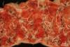
Description: The wall of the large intestine was severely edematous and hyperemic. The content was watery admixed with large amounts of mostly uncoagulated blood. Additionally, high numbers of 20-50 mm long, beige nematodes were found. Parasites were mostly attached to the mucosa with their <1 mm thin anterior end and presented a 1-2 mm thick, often curled posterior end. The skin of the anal region was smeared with blood and feces.
Comment: The Baboon presented clinically with cachexia that had been mainly attributed to high age and intermittent endoparasite infestation. After her spontaneous death she was submitted to pathological examination. Macro- and microscopic examination revealed a severe, hemorrhagic typhlocolitis as well as large amounts of trichuris-type nematodes. Bacterial culture revealed a mixed culture.
Trichuris spp. are extremely common parasites of the intestinal tract of free ranging and captive baboons. Their pathology ranges from subclinical infection to severe enteritis. Trichuriasis in the baboon represents a zoonosis, especially in free ranging baboons foraging human waste. In humans it is considered a neglected tropical disease and affects roughly 600 million people worldwide. Trichuris spp. are also common in other primates, ruminants, pigs as well as dogs and occur in others species including mice, lagomorphs and cats. They have not been found in the horse.
Picture and Authored by Moritz Radbruch, Department of Veterinary Pathology, Freie Universitaet Berlin, Germany