Animal: Sea Clown Surgeon (Acanthurus sohal), juvenile, sex unknown
Organ: Gill
History: Death during quarantine. Hyperactive prior to death.
Necropsy findings: Numerous slightly raised, 0.5-1mm, grayish to white nodules on gills and skin accompanied by moderate amounts of mucous.
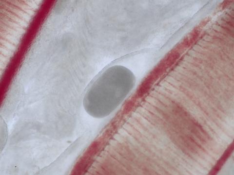
Cytology findings: The branchial epithelium is locally raised and hyperplastic, encysting a large subepithelial, oval, indistinctly ciliated parasite with abundant, finely granular to vacuolated cytoplasm and a macronucleus (partially visible, appears oval shaped in image, video demonstrates linked, bead-like segments) (trophont). The organism exhibits characteristic ‘rolling’ movement (video).
Morphological diagnosis: Gill, epithelial hyperplasia, nodular, moderate with subepithelial protozoal trophont consistent with Cryptocaryon irritans
Ethiological diagnosis: Branchial Cryptocaryonosis
Ethiology: Cryptocaryon irritans
Name of the disease: Marine Ich / Marine White Spot Disease
Comment: This gill snip of a freshly dead fish demonstrates infection with Cryptocaryon irritans, the causative agent of Marine Ich. This parasite has long been referred to as the marine / saltwater ‘counterpart’ of Ichthyophthirius multifilis, the causative agent of Freshwater white spot disease (Ichthyophthiriosis). Whilst both organisms have the same life cycle and pathology, this was shown to be due to convergent evolution rather than phylogenetic relatedness. Affected skin often appears like it was finely dusted with salt. Virtually any teleost is probably susceptible. The life cycle is 11-15 days but can be completed in as little as 6 days at optimum reproduction temperature. In both Cryptocaryon and Ichthyophthirius, adult ciliated protozoa (theronts – free-swimming, non-feeding, infective stage) adhere to the epithelium of gills or skin and burrow into the epithelium. This results in nodular epithelial hyperplasia around the trophont (host fish-feeding stage). Trophonts erupt through the epithelium, drop off the host, and form a capsule (tomont – encysted, dividing stage), adhering in environment. Following binary fission this produces hundreds to thousands of tomites (daughter cells produced by a tomont) which become infective, motile theronts, completing the life cycle. Detection of an oval, holotrich (evenly dispersed cilia over entire body) ciliated parasite encysted within host’s epithelium is pathognomonic. Whilst Ichthyophthirius presents with a C-shaped macronucleus, the macronucleus of Cryptocaryon is moniliform, consisting of four linked, bead – like segments. Traditionally a problem in aquarium fish, Cryptocaryon irritans has become a serious disease in cultured warm water marine food fish and has caused epidemics in wild marine fish.