History: 13-years-old, male, racoon, Procyon lotor
Diagnosis: Unilateral thyroid adenocarcinoma and maxillary (gingival) squamous cell carcinoma
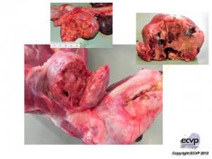
Description: At necropsy, the left maxilla was severely thickened by a approximately 3.2×2.8×2 cm in size, infiltrative growing, and poorly demarcated neoplasia which on the cut surface was whitish. The left thyroid was markedly enlarged, approximately 6.5x6x4.5 cm in size and on the cut surface was cystic and contained necrotic material.
Comments: This was an interesting case of an unusual presentation of three neoplasms – thyroid adenocarcinoma, maxillary squamous cell carcinoma and pheochromocytoma – in a 13-years-old raccoon from a zoo in Berne. The patient was euthanatized because of the poor body condition. At necropsy, three neoplastic lesions were observed at the level of the left thyroid, left part of the maxilla and right adrenal gland. In histology, the maxillary mass consisted of nests of epithelial cells differentiated into squamous epithelial with “keratin pearls” formation which are typical features of squamous epithelial carcinoma. The thyroid mass showed tubular, sometimes papillary, epithelial structures filled with eosinophilic homogenous material (colloid), as well as cystic dilations of the follicles and scant fibrous stroma. The adrenal mass was histopathologically diagnosed as pheochromocytoma.
Picture by: Francesca Popescu
Author: Francesca Popescu, Dominique Wiener, Institute of Animal Pathology, University of Bern, Vetsuisse Faculty, Switzerland