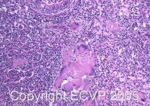
Cattle lung: Bronchointerstitial pneumonia with formation of multinucleate syncytial cells
Detailed information
Animal: Calf, weight: 65 kg, Norwegian red cattle.
Organ: Lung
History: Chronic cough, abnormal respiratory sounds. Treated with antibiotics 7 weeks prior to autopsy. No other clinical signs. Similar signs in other calves in the herd. This animal was submitted for autopsy for diagnostic purposes.
Autopsy findings: Bilateral cranioventral consolidation of the lungs. In the consolidated tissue, large amounts of neutrophils were found in the lumina of bronchi, bronchioli and alveoli, as well as in the interstitium. The interstitial purulent exudate was mixed with lymphocytes, and macrophages. Numerous multinucleate syncytial cells were seen in the inflamed lung tissue. Proliferation of type II alveolar epithelial cells was present. Acid fast staining for mycobacteria (Ziehl-Neelsen) was negative. Cultivation from mediastinal lymph node and lung tissue yielded Pasteurella multocida in abundance.
Diagnosis: Lung, pneumonia, bronchointerstitial, with formation of syncytial cells and secondary purulent bronchopneumonia. The lesion was deemed to be consistent with paramyxovirus infection, (probably bovine respiratory syncytial virus) with secondary Pasteurella multocida infection.
Necropsy performed by: Dr. G. Gunnes.
Photo by: Dr. G. Gunnes.