Malignant amelanotic melanoma, oral mucosa, dog
Detailed information
Animal: Canine, giant schnauzer (salt&pepper), 9 years, male.
Organ: Oral biopsy.
History: The dog developed av grayish, firm, multilobulated tumour on the inside of the upper lip.
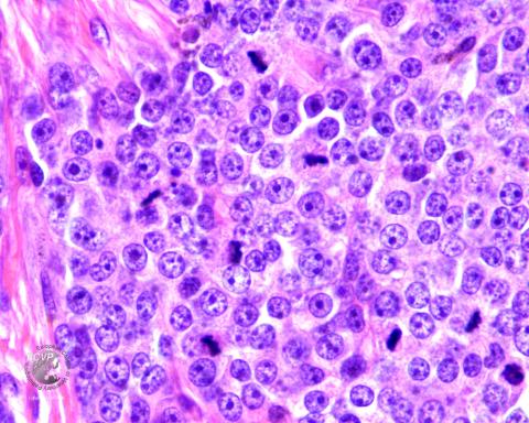
Biopsy findings: A cell rich tumour tissue consisting of large, poorly differentiated cells with large, mainly rounded nuclei was found. The nuclei contained one to several, very distinct nucleoli, which in some cases were enlarged up to the size of erythrocytes. The eosinophilic cytoplasm was moderate and the cell boundaries were usually distinct, and round to edgy. Scattered melanomacrophages with numerous, large melanin granules were found throughout the tissue. A large number of mitoses were found and up to 15 were counted in a 400x field. The cells invadede aggressively in surrounding connective tissue.
Diagnosis: Malignant amelanotic melanoma, oral mucosa, dog.
Photo by: Gjermund Gunnes