History: Cat, European shorthair, 12 years, neutered male.
Diagnosis: Duodenum: Feline gastrointestinal eosinophilic sclerosing fibroplasia
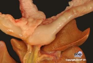
Description: A firm mass of 2.5 x 1.5 x 1.5 cm expanded the duodenum at the level of the duodenal papilla. Within the mass, the mucosa was ulcerated and the duodenal lumen was almost completely obstructed. In addition, large areas of the small intestine were filled with coagulated blood.
Comments: The cat was clinically presented with hematemesis and severe progressive anaemia for one week and was euthanized due to poor prognosis (final haematocrit 12%). The main lesion at necropsy was a duodenal mass. Macroscopic differentials to be considered in this location in cats include intestinal lymphoma, intestinal adenocarcinoma, intestinal mast cell tumour and feline gastrointestinal eosinophilic sclerosing fibroplasia. Histology revealed a mainly eosinophilic and less lymphoplasmacytic inflammatory infiltrate separated by large reactive fibroblasts mostly surrounding small blood vessels and branching and anastomosing trabeculae of dense collagen. An eosinophilic inflammation and fibroplasia was also found in several lymph nodes (e. g. mesenteric, mandibular, popliteal). Sclerosing mast cell tumour and duodenal osteosarcoma have been described as histologic differentials. However, the few descriptions of the latter have been discussed to be misdiagnoses of feline gastrointestinal eosinophilic sclerosing fibroplasia.
Picture by: Anja Ostrowski;
Author: Sophie Bader ;Department of Veterinary Pathology, Freie Universität Berlin, Germany