Animal: 8-year-old, neutered male, Siamese, cat
Organ: Subcutis
History: One week before initial presentation, owners noticed a right subcutaneous inguinal mass. Clinicians described a non-painful, non-ulcerated, subcutaneous, 12 x 7 cm mass. A fibrosarcoma was suspected and a fine-needle aspiration was performed but was inconclusive (slides only contained blood). Radiographs showed multiple pulmonary metastases. A palliative treatment was initiated. Two months later, the animal was presented for anorexia and prostration, and euthanasia was elected. At this time, the mass reached 15 x 10 cm. A necropsy was performed.
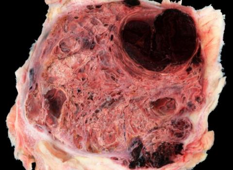
Gross findings: At necropsy, the animal was cachectic. The right inguinal area contained a well-demarcated, encapsulated, 12 x 9 x 9 cm mass. Cut section revealed a red, cystic and spongy parenchyma with blood clots. The lungs contained multiple 1-5 mm dark red nodules highly suggestive of metastatic hemangiosarcoma. A 50 ml hemothorax was also noted.
Histopathological findings: Histological examination of the mass showed a typical hemangiosarcoma and pulmonary metastases were confirmed.
Diagnosis: Subcutaneous hemangiosarcoma with pulmonary metastases and hemothorax.
Comment: The incidence of hemangiosarcoma is lower in the cat than in the dog and there are few studies on feline cutaneous/subcutaneous hemangiosarcomas. A good long-term prognosis is possible for cats treated with aggressive surgical excision. In this species, hemangiosarcoma also affect the head, pinnae, conjunctiva and cornea, and a causal association with UV exposure is highly suspected at these sites.
In the absence of metastases, gross diagnosis of hemangiosarcoma can be difficult, especially in the spleen of dogs where hematoma and nodular hyperplasia are the main differential diagnoses.
Other vascular proliferations/neoplasms that should be known in the cat are the so-called plexiform vasculopathy/vascularization of lymph nodes that can transform into nodal hemangiosarcoma; feline abdominal ventral lymphangiosarcoma (formerly known as angiosarcoma); and feline systemic reactive angioendotheliomatosis.
Reference:
FUJI RN., PATTON KM., STEINBACH TJ., SCHULMAN FY., BRADLEY GA., BROWN TT., et al. Feline systemic reactive angioendotheliomatosis: eight cases and literature review. Vet Pathol. 2005, 42, 608 617.
HENDRICK MJ. Mesenchymal tumors of the skin and soft tissues, in: Tumors in Domestic Animals. 2017, editor: Donald J. Meuten, Ames, Iowa, p. 142 175.
JUNGWIRTH N., JUNGINGER J., ANDRIJCZUK C., BAUMGÄRTNER W., WOHLSEIN P. Plexiform Vasculopathy in Feline Cervical Lymph Nodes. Vet Pathol. 2018, 55, 453 456.
MCABEE KP., LUDWIG LL., BERGMAN PJ., NEWMAN SJ. Feline cutaneous hemangiosarcoma: a retrospective study of 18 cases (1998-2003). J Am Anim Hosp Assoc. 2005, 41, 110 116.
PIRIE CG., DUBIELZIG RR. Feline conjunctival hemangioma and hemangiosarcoma: a retrospective evaluation of eight cases (1993-2004). Vet Ophthalmol. 2006, 9, 227 231.
ROOF-WAGES E., SPANGLER T., SPANGLER WL., SIEDLECKI CT. Histology and clinical outcome of benign and malignant vascular lesions primary to feline cervical lymph nodes. Vet Pathol. 2015, 52, 331 337.
SHANK AMM., TEIXERIA LBC., DUBIELZIG RR. Canine, feline, and equine corneal vascular neoplasia: A retrospective study (2007-2015). Vet Ophthalmol. 2019, 22, 76 87.
Photo (after water immersion of the specimen) by: Edouard REYES-GOMEZ, Unité d’Histologie et d’Anatomie Pathologique, Ecole Nationale Vétérinaire d’Alfort, France
Author: Edouard REYES-GOMEZ, Unité d’Histologie et d’Anatomie Pathologique, Ecole Nationale Vétérinaire d’Alfort, FRANCE