Cattle brain: Malignant catarrhal fever
Detailed information
Animal: Cow, Norwegian red cattle
Organ: Brain
History: 2 year old bull, small for its age. Increased heart and respiratory rate, reduced appetite, dehydrated. Multiple skin vesicles with some exudation. Impaired sight, periocular swellings, conjunctivitis, corneal edema. Oral erosions, enlarged prescapular lymph nodes.
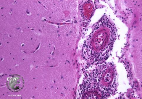
Autopsy findings (excerpt): Multiple vesicular papules and erosions in the skin. Lymph nodes were considerably enlarged. The cornea was clouded bilaterally. Diffuse, coalescing erosions were found on the tounge and multifocally in the tracheal and esophageal mucosae as well as in the abomasum. A distinct histological finding in all examined organs was a marked necrotising vasculitis and perivascular infiltration, predominantly by lymphocytes. In all segments of the brain and meninges, blood vessel walls and perivascular spaces were infiltrated by lymphocytes. In the eye, a profuse infiltration of mononuclear inflammatory cells was seen in the iridocorneal angle, iris, sclera and pervascularly around the optic nerve.
Diagnosis: Cow, Brain, Malignant catarrhal fever
Necropsy performed by: Caroline Piercy Åkesson
Photo by: Gjermund Gunnes