Signalement: Horse, Paint Horse, 1 year old, female.
History: The horse presented with acute onset of right-forelimb lameness. Aggravated clinical signs, radiographically evident sinking and rotation of the distal phalanx and additional acute affection of the left forelimb resulted in poor prognosis and subsequent euthanasia.
Diagnoses:
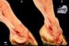
A: Left forelimb, phalanx distalis: acute, diffuse, moderate laminitis
B: Right forelimb, phalanx distalis: chronic, diffuse, severe laminitis with sinking and rotation of the distal phalanx and compression atrophy of adjacent hoof structures?
Description: A: Affecting the distal phalanx of the left forelimb, there was a diffuse, moderate hyperemia of the corium and the tip of the distal phalanx. There was marked thickening of the corium surrounding the distal phalanx.
B: The distal phalanx of the right forelimb had diffuse, severe hyperemia of the surrounding corium and bone with marked sinking and rotation of the phalanx and deformation of the hoof wall, resembling chronic lamintis.
Comments: The distal phalanx is held by the interdigitation of the epidermal lamina (inner hoof wall) and the corium which is attached to the phalanx. If the loss of adhesion between the epidermal and dermal laminae is sufficient and the forces of weight and the deep flexor tendon exert additional mechanical stress, tearing of the remaining laminae and rotation or distal displacement of the distal phalanx may result.
Systemic diseases that result in laminitis include either severe inflammation (e.g. gastrointestinal disease, endometritis leading to sepsis, carbohydrate overload, exposure to black walnut extract) or endocrinopathies (e.g. equine metabolic syndrome with hyperinsulinemia and insulin resistance or pituitary pars intermedia dysfunction PPID with ACTH overproduction). Laminitis may also occur as a consequence of contralateral limb overload (most likely in this case at the left forelimb).
The pathogenesis of laminitis is under intense investigation and seems to vary depending on the primary cause. For example, inflammatory variants probably induce a systemic inflammatory response syndrome (SIRS) with activation of matrix metalloproteinase and leukocyte infiltration. On the other hand, endocrinopathies might lead to lamellar failure based on insulin mediated disturbances in the epidermal – dermal metabolism and structure.
Acute laminitis presents with sudden lameness and severe pain, increase in the hoof wall temperature and a digital pulse. Gross lesions are limited to congestion of the laminar dermis and occasionally hemorrhage without hoof deformity (see figure A). The skin above the coronary band might be swollen, indicating acute separation and displacement of the phalanx distalis (“sinkers”). Early histologic lesion including elongation and disorganization of the basal keratinocytes and attenuation of the secondary epidermal laminae. The detachment of the basal membrane of secondary laminae, the collapse of the secondary laminae and the loss of capillaries finally leads to loss of structural integrity of the phalanx and hoof wall due to ischemic damage.
Chronic laminitis is presented with several days of lameness or when rotation of the distal phalanx is evident (see figure B). Relevant factors for the rotation include body weight, the leverage forces placed on the toe and the pulling forces of the deep digital flexor tendon. At the worst, penetration of the hoof’s sole is possible. Histology reveals marked irregular hyperplasia of the epidermal laminae with hyperkeratosis.
Picture and authored by: Angele Breithaupt, Department of Veterinary Pathology, Freie Universitaet Berlin, Germany