Diagnosis: Striped dolphin. Lung. Severe, multifocal, chronic, bronchointerstitial pneumonia. Moderate, multifocal, subacute, pyogranulomatous bronchopneumonia with intralesional nematodes.
Animal: Adult, male, striped dolphin.
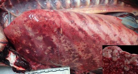
Organ: Thoracic cavity and lung.
History: The animal stranded alive with neurological signs, died soon after.
Gross findings: The lung appear expanded with ribs impression and multiple darks slightly depressed areas (atelectasis). Affecting approximately the 20% of the lung parenchyma there are multifocal, well delimitated, raised, firm, tan to white lesions, that frequently track the small caliber bronchi.
Histopathological findings: Multifocally the lung interstitium is markedly infiltrated by lymphocytes and histiocytes. The lumens of the alveoli and airways are frequently occluded by large amount of histocytes and type II pneumocytes. Syncytia are frequently observed within the alveolar spaces. There are both intracytoplasmic and intranuclear, acidophilic, small inclusion bodies of about 1 to 3 micrometers. Multifocally in the bronchi there are nematodes and numerous larvae surrounded by moderate pyogranulomatous inflammation.
Etiological diagnosis:
Morbilliviral pneumonia.
Verminous pneumonia.
Etiology:
Cetacean morbillivirus (confirmed by IHQ)
Halocercus sp.
Investigation and photo by: Cristian M. Suárez Santana, Antonio Fernández. Institute for Animal Health and Food Safety. University of Las Palmas de Gran Canaria.