Diagnosis: Cat, skin: Dermatitis, deep, nodular, granulomatous, marked, with intralesional, intracytoplasmic yeasts (consistent with Histoplasma sp.).
Animal: 1-year-old, female neutered, European shorthair
History: Dome-shaped mass in the area of the left hind foot, 5th toe. Surface looked like scalped. No history of travelling. Clindamycin treatment but became thicker. Surgical excision: wound healed well.
Histological findings: Histopathologic evaluation of sections of the nodule revealed on low power a nodular dermatitis that extended into the deep subcutis. The overlying epidermis of the sample was focally extensive ulcerated. From the ulcerated epidermis/subepidermal dermis up to the deep subcutis, there were infiltrated of numerous epithelioid macrophages intermingled with less plasma cells, lesser lymphocytes and neutrophils. At higher magnification, macrophages were commonly distended by up to 20 intracytoplasmic yeast bodies that were round, 3 to 4 µm diameter and had a basophilic center surrounded by a 2 µm halo (consistent with Histoplasma sp.).
Periodic acid Schiff stain (PAS) stain showed pink intracytoplasmic yeasts.
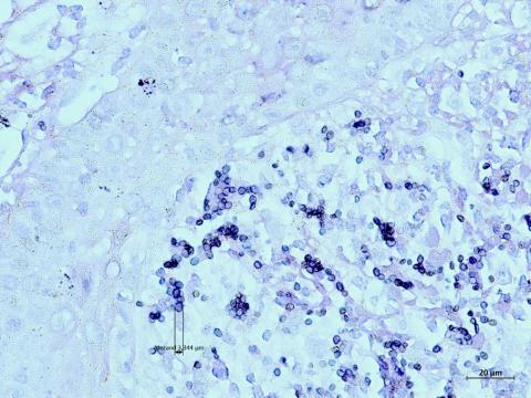
GMS stain showed fungal intracytoplasmic black-colored yeasts.
For a definitive diagnosis and species identification, molecular biology has been used. After extraction from the paraffin block, DNA was amplified by Multilocus Sequence Typing and by specific qPCR.
Authors and photos by: Dr. Stephanie Muller, Dipl. ECVP, Idexx Vet Med Labor GmbH, Dr. Ursula Mayer, Dipl. ECVD, Idexx Vet Med Labor GmbH, Dr. Dunja Wilmes, Robert-Koch-Institut, Berlin