Animal: Dog, mixed breed, 8 years old
Organ: Rectum (segmental section)
History: This case was initially described as a rectal polyp in a dog and subsequently excised for histopathological evaluation. A firm intramural mass was detected during abdominal ultrasonography.
Aetiology: Reactive nodular hyperplasia of lymphoglandular complexes.
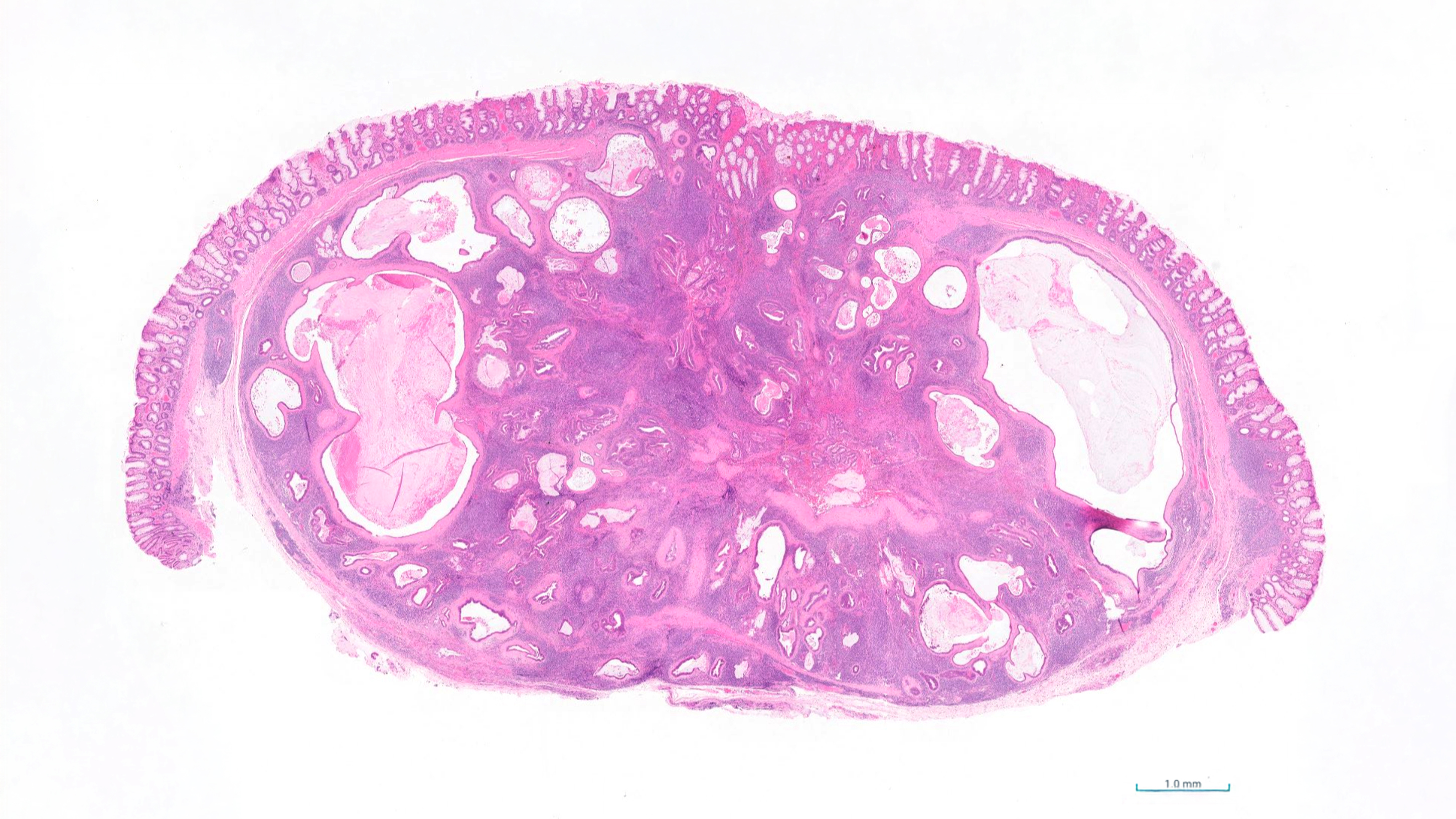
Macroscopic findings: The rectal wall contained a well-demarcated, firm, pale tan nodule measuring approximately 1.5 cm in diameter. The overlying mucosa was intact and smooth. On cut section, the lesion was homogeneous and lacked evidence of necrosis or cavitation.
Morphological diagnosis: Rectum: focal, well-demarcated, chronic nodular hyperplasia of lymphoglandular complexes.
Histopathology: The lesion consisted of well-organised lymphoid aggregates and follicles intimately associated with glandular profiles, some distended with mucus. No significant dysplasia, cellular atypia, or evidence of malignancy was observed. The architecture was consistent with a reactive process rather than neoplastic transformation.
Comment: Nodular hyperplasia of lymphoglandular complexes in the rectum of dogs is an uncommon but recognised benign condition characterised by focal proliferation of lymphoid and glandular tissue within the rectal mucosa and submucosa.
Lymphoglandular complexes are components of the gut-associated lymphoid tissue, formed by a close association between lymphoid follicular tissue and epithelial diverticula projecting from the overlying mucosa. These structures are predominantly located within the distal small intestine, caecum, and colon and have been documented in a wide range of mammalian species, including humans, dogs, cattle, horses, pigs, dolphins, and echidnas.
Hyperplastic lesions of lymphoglandular complexes are rarely identified histologically. The incorporation of an epithelial component into these lesions introduces a potential risk of misdiagnosis as either carcinoma or lymphoma.
The process is considered reactive rather than neoplastic, typically associated with chronic antigenic stimulation or local inflammation. Recognition of this entity is important to avoid misdiagnosis of lymphoma or adenocarcinoma.
Interestingly, similar lesions have been well documented in humans (often termed inverted lymphoglandular polyps), with a seemingly higher frequency of presentation—likely attributable to the more extensive use of advanced imaging techniques. In people, these nodules appear to be asymptomatic or associated with mild clinical signs such as rectal bleeding or discomfort, and they are generally regarded as benign with no significant risk of progression to lymphoma.
Photo prepared and performed by: Richard Fox
Bibliography:
Stent AW, Kiupel M, Dandrieux JRS, Liffman R, Bera MM. Nodular hyperplasia of lymphoglandular complexes in dogs: A potential diagnostic pitfall for rectal masses. Veterinary Pathology. 2023;61(2):243-247. doi:10.1177/03009858231190643
Zhou, Shengmei, Ma, Yanling, Chandrasoma, Parakrama, Inverted Lymphoglandular Polyp in Descending Colon, Case Reports in Pathology, 2015, 646270, 3 pages, 2015.