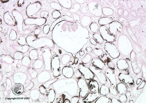
Dog, kidney: Hypercalcemic nephropathy, von Kossa Stain
Detailed information
Animal: 5 months, airdale terrier, male.Organ: Kidney
History: Hemorrhagic diarrhoea and vomitus. Blood samples indicated kidney failure. The clinical signs had a relatively sudden onset.
Histological findings: The cortical interstitium showed oedema and a moderate amount of plasma cells and fibroblasts with an increase in distance between tubular structures. A number of tubules and capsular spaces were dilated. There were streaks of connective tissue extending from the capsule through the cortex towards the medulla. An increased amount of immature nephrons were found, often associated with the cortical scars. The tissue had extensive calcium deposits in tubular basement membranes, Bowman’s capsule and vessel walls. This was not apparent in the H&E stain, but was easily detected in the von Kossa stain for calcium.
Diagnosis: Hypercalcemic nephropathy, unknown aetiology
Histology performed by: Gjermund Gunnes
Photo by: Gjermund Gunnes