Animal: a 11-year-old, neutered female, Labrador Retriever dog.
Organ: cervical spinal cord.
History: nonambulatory paresia of the hindlegs and the left foreleg, with a 3-day history of loss of proprioception. Presence of left-sided Horner’s syndrome.
Autopsy (performed by Laëtitia Dorso): absence of significant gross lesion.
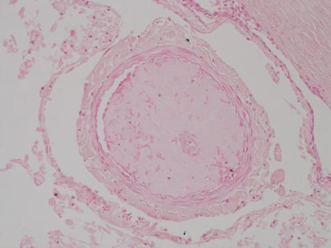
Histopathological diagnosis: polio- and leucomyelomalacia, focally extensive, severe, with wallerian degeneration, haemorrhage and fibrocartilagenous emboli.
Potential additional stainings to highlight the emboli: Alcian blue or Toluidine blue.
Name of the condition: fibrocartilaginous embolic myelopathy.
Histopathology and photo by: Cynthia Robveille, Autopsy service, Oniris – Veterinary Medicine School, Nantes, France.