Animal: Ovine (Ovis aries), Merina breed, female, 7 years old.
Organ: Liver
History: Animal from an extensive breeding farm with non-diarrheic, chronic progressive wasting affecting animals from one year and beyond. This case presented mixed dyspnea, strong productive cough and severe anemia. Anaplasma ovis was diagnosed by PCR from blood samples and treated. Echography revealed multiple anecoic cysts in the liver. The animal did not respond to treatment and it was euthanized.
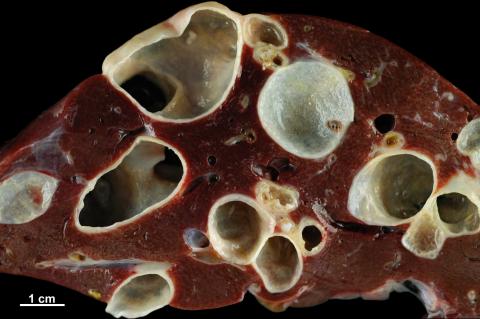
Etiology: Echinococcus granulosus
Macroscopic findings: Liver is altered by the formation of multiple 1 to 2.5 cm turgid cystic and expansive cavities, some of them embedded in the hepatic parenchyma and others protruding on the surface. Cut section demonstrated empty cavities delimited by a thickened wall. The internal surface of the cavities was irregular and granular, showing a whitish stippling compatible with protoscolices attached to the internal face of the wall.
Morphological diagnosis: Liver: Multifocal, unilocular hydatid cysts with parenchymal compression.
Comment: Further necropsy findings included a single hydatid cyst in the right apical lung lobe. Histological findings revealed representative Echinococcus granulosus intermediate stages, with unilocular cysts delimited by an inner cellular germinal layer with protoscolices together with a laminated layer and an outer fibrous adventitial layer of the host.
Necropsy performed by: Estela Pérez*, Álex Gómez, Ana Rodríguez, Raúl Reséndiz, Ricardo de Miguel and Lluís Luján. Department of Animal Pathology, Veterinary Faculty, University of Zaragoza, Spain.
Photo prepared and performed by: Estela Pérez, Álex Gómez and Ana Rodríguez.