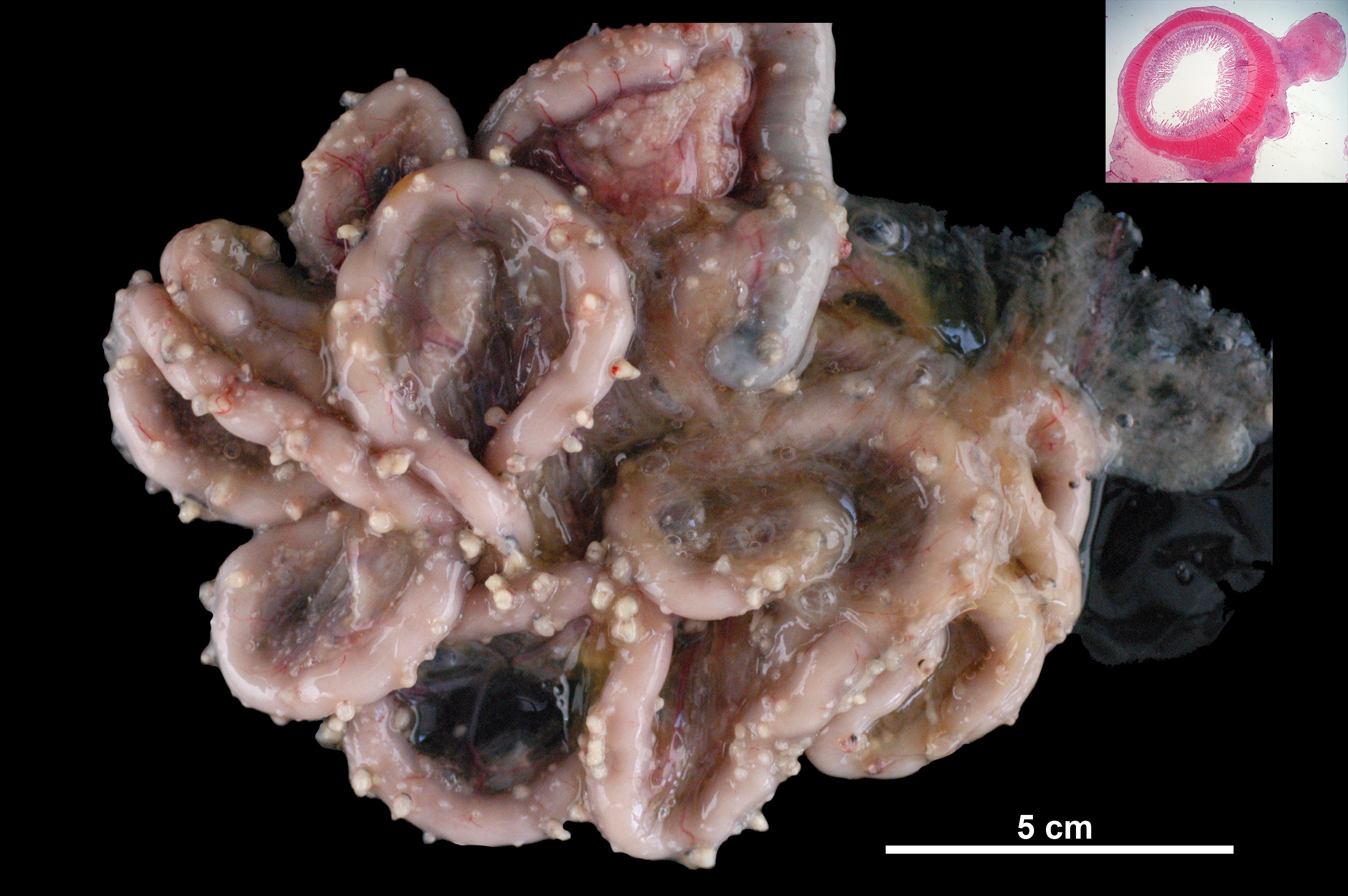
Animal: Cat, European Shorthair, neutered male, 10.5 years old
Organ: Intestinal tract
History: Chronic wasting for 7 months and abdominal distension for four weeks. The animal showed nervous signs and mydriasis. Decreased serum albumin and increased serum globulins. Peritoneal fluid was protein-rich (Rivalta test positive).
Etiology: Feline infectious peritonitis virus (Alphacoronavirus).
Macroscopic findings: Multifocally, the serosa of both, small and large intestine is expanded by up to 0.5 cm whitish, protruding nodular proliferations. There is severe edema in the mesentery.
Morphological diagnosis: Intestinal tract (serosa): Severe, chronic, multifocal pyogranulomatous peritonitis.
Comment: In cases of FIP, formation of exophytic pyogranulomatous inflammatory nodules in the intestinal serosa is an unusual event. Two complementary microscopic images are included.
Necropsy performed by: Álex Gómez, Natalia Calvo and Marcelo de las Heras, Department of Animal Pathology, Veterinary Faculty, University of Zaragoza, Spain.
Photo prepared and performed by: Álex Gómez*, Natalia Calvo, Estela Pérez and Lluís Luján.
* a.gomez@unizar.es