Animal: Dog, mixed-breed, neutered male, 6.5-years-old
Organ: Brain.
History: The dog was presented with a history of refractory seizure activity. He was diagnosed with a left parietal brain mass and underwent radiation therapy. After 4 months, he had an increase in seizures severity and frequency, and MRI showed that the tumor doubled in size. The dog was humanely euthanized and presented for autopsy at the diagnostic laboratory of the Cummings School of Veterinary Medicine.
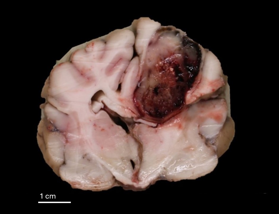
Macroscopical findings: In the parietal lobe of the cerebral left hemisphere, there was an intraparenchymal nodular mass, about 2.5x2.5x1cm large, which was moderately demarcated, firm, tan- to greyish, with hemorrhages. The mass compressed the adjacent parenchyma, caused a deviation of the midline, and slightly bulged on the surface of the brain.
Morphological diagnosis: High-grade astrocytoma (glioblastoma)
Contributor: Laura Nordio, DVM, PhD, DECVP. Cummings School of Veterinary Medicine at Tufts University, North Grafton, MA, USA (current affiliation: MYLAV La Vallonea Veterinary Diagnostic Laboratory, Italy)