Text
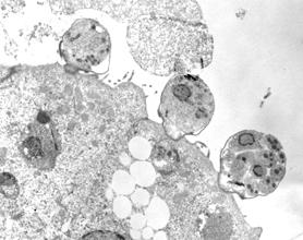
TEM micrograph of trofozoites and meront stages in a Cryptosporidim scophtalmi n. sp. infection in turbot (Psetta maxima)
Detailed information
History: From 1997 to 2000 a parasitological survey was carried out in different turbot farms in NW Spain involving more than 8000 fish.
Diagnosis: TEM micrograph of trofozoites and meront stages in a Cryptosporidim scophtalmi n. sp. infection in turbot (Psetta maxima).
Autopsy findings: Intestinal distension arising from mucous intestinal contents and gas was observed in some fish. Other coelomic viscera were normal. In Giemsa-stained mucosal scrapings ooquist showing metachromasia were detected.
Photo by: Maria Isabel Quiroga (author), José María Nieto (editor)