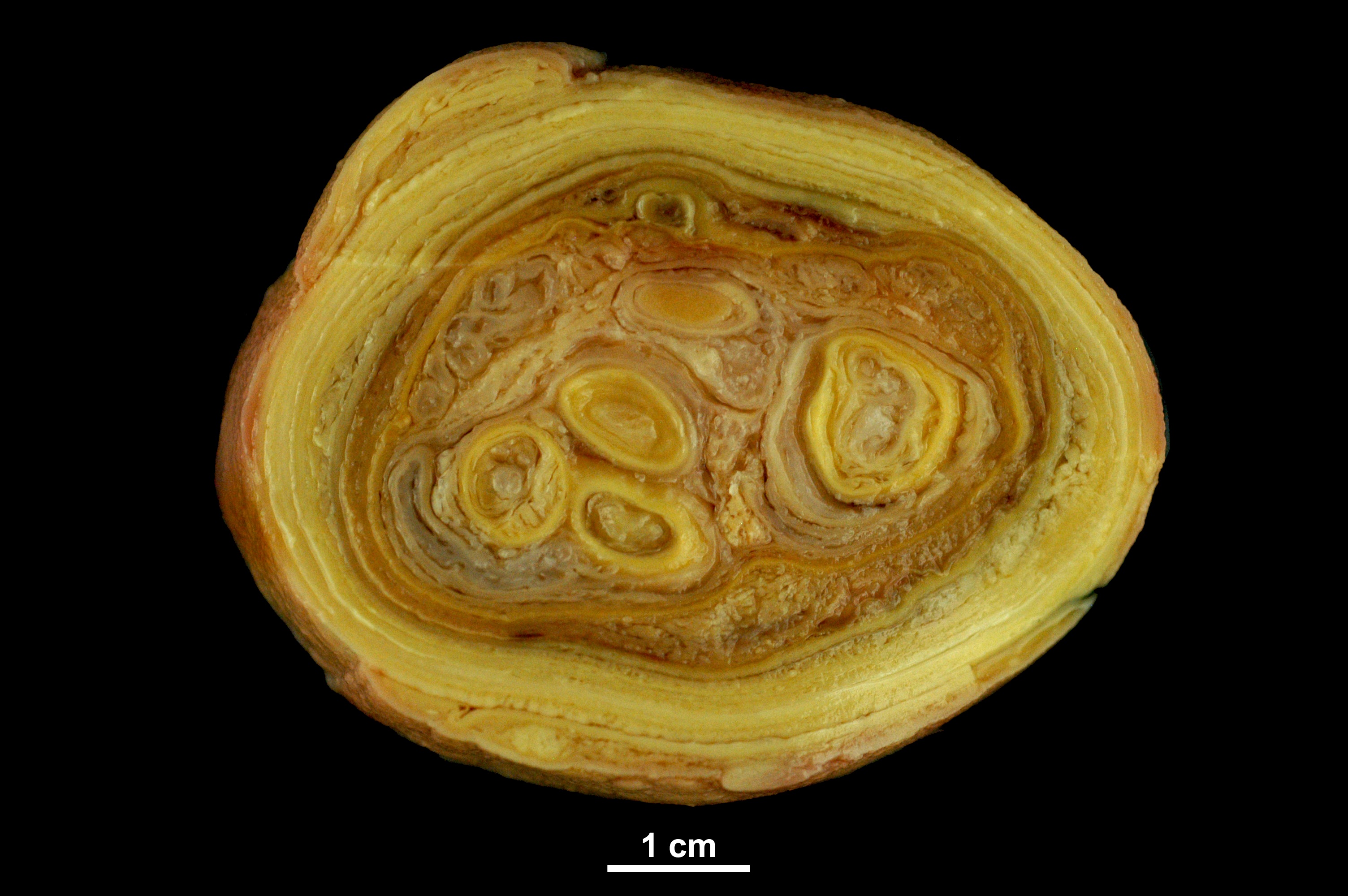
Animal: Backyard laying hen, unknown age.
Organ: Oviduct (transversal section)
History: The animal egg-laying declined sharply before its sudden death.
Etiology: Escherichia coli (most common). Rarely Gallibacterium anatis.
Macroscopic findings: The oviduct was distended showing thin walls and a firm consistency. Upon sectioning, it contained an obstructive plug of yellowish-white caseous material arranged in multiple concentric layers.
Morphological diagnosis: Oviduct: focal extensive, severe, chronic, caseous fibrinoheterophilic salpingitis.
Comment: The multilayered appearance results from alternating layers of fibrinoheterophilic exudate (looser, lighter layers) and coagulated ova and albumin secretions (denser layers). Over time, the exudate accumulates and mixes with cyclically produced egg secretions following each ovulation.
Necropsy performed by: Estela Pérez*, Álex Gómez, Alicia de Diego and Lluís Luján. Department of Animal Pathology, Veterinary Faculty, University of Zaragoza, Spain.
Photo prepared and performed by: Lluís Luján, Estela Pérez and Álex Gómez.
Bibliography:
Abdul-Aziz T, Barnes JH. Bacterial peritonitis, salpingitis, and salpingoperitonitis. In: Gross Pathology of Avian Diseases, Text and Atlas. Jacksonville, Florida: The American Association of Avian Pathologists, Inc.; 2018:35–38