Dog: Oligodendroglioma with atrophy of adjacent brain parenchyma
Animal: Dog, Husky, 10 years, female
Diagnosis: Cerebrum, left hemisphere: Oligodendroglioma with atrophy of adjacent brain parenchyma
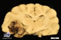
Description: A well demarcated, dark grey to brownish mass with maximal extension of 1.5 cm in diameter was present in the left temporal lobe, compressing and replacing surrounding neuropil.
Comments: The dog presented clinically with sudden onset of circling behavior followed by unilateral facial paralysis and death. Necropsy revealed scull asymmetry and a greyish-brownish mass in the cerebrum which presented histologically as an oligodendroglioma with areas of necrosis, replacing and compressing surrounding tissue. There was no evidence of metastatic spread. In addition to the brain lesion there were mild, chronic, multifocal, lymphoplasmacytic pyelitis and interstitial nephritis as well as mild, acute, multifocal, myocardial hemorrhages.
Oligodendrogliomas are relatively common primary neoplasms of the central nervous system in dogs. There is a certain predisposition in brachycephalic breeds and the incidence increases with age. Usually the frontal, parietal or temporal lobes are affected. At necropsy oligodendrogliomas often appear dark due to multifocal hemorrhages and large tumors may have central cystic change.
Picture and authored by: Aleksandra Żuraw, Department of Veterinary Pathology, Freie Universität Berlin, Germany