Animal: Dog, mixed-breed, female, 13-years-old
Organ: Haired skin
History: The dog had a paravaginal subcutaneous mass.
Macroscopical findings: Sample of skin and subcutis with a 3-cm-large soft, whitish, multilobular subcutaneous neoformation.
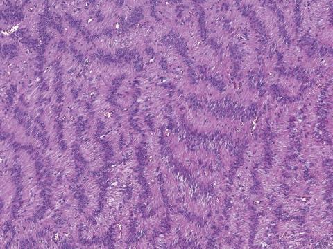
Histopathological findings: Subcutaneous infiltrating multilobular neoplasm, from densely (“Antoni A” areas) to sparsely (“Antoni B” areas) cellular, organized in short bundles and frequent Verocay bodies. Neoplastic cells are spindle, 8-10 μm in transverse diameter, with intermediate N/C ratio, indistinct cell borders, moderate amount of eosinophilic fibrillar cytoplasm, paracentric oval nuclei with dispersed chromatin and small nucleolus. Moderate anisocytosis and anisocariosis. 0-2 mitoses/HPF. H&E stain.
Diagnosis: Haired skin. Malignant peripheral nerve sheath tumor (MPNST)
Contributor: Laura Nordio, San Marco Veterinary Clinic and Laboratory, Veggiano (PD), Italy