Species and history: Dog, 2 yrs old, female neutered
Circular cutaneous mass excised from the abdomen, purple in color with a pointed center, approximately 1cm in diameter.
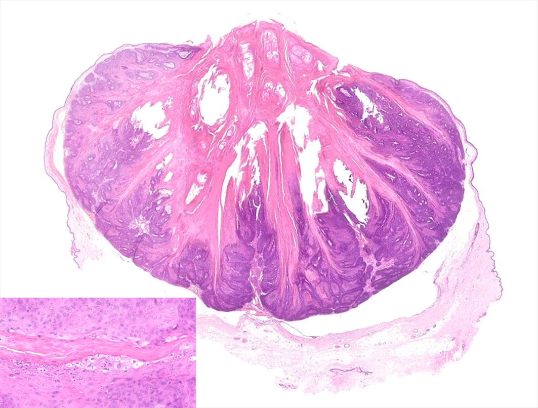
Organ: Skin
Description:
Within the dermis there is an expansile cup shaped inverted papilloma composed of thick squamous epithelium. Epithelial fronds are heavily keratinized and forming spikes. This hyperplastic epithelium exhibits extensive evidence of viral cytopathic effect, including the presence of numerous koilocytes, hypergranulosis with giant keratohyalin granules, and occasional ground-glass intranuclear inclusion bodies with peripheralized chromatin.
Arising from the epidermis and expanding the dermis is a well-demarcated, crateriform, endophytic neoplasm composed of squamous epithelial cells progressing from a hyperplastic stratum basale to a thickened stratum spinosum (acanthosis) and stratum granulosum and forming papillary projections into a central keratin-filled cavity. Neoplastic cells have distinct cell borders, a moderate amount of eosinophilic cytoplasm, and round to oval nuclei with finely stippled chromatin and 1-4 prominent magenta nucleoli. The stratum spinosum contains scattered keratinocytes with pale, swollen, vacuolated cytoplasm and pyknotic nuclei occasionally surrounded by a clear halo (koilocytes). The stratum granulosum frequently contains large irregular intracytoplasmic keratohyalin granules. There is prominent orthokeratotic and parakeratotic hyperkeratosis and few multifocal aggregates of ghost cells. Within the adjacent compressed dermis, there is a diffuse infiltrate of moderate numbers of lymphocytes, plasma cells, and fewer macrophages with few small areas of hemorrhage.
Morphological diagnosis: Cutaneous interted papilloma
Etiologic diagnosis: Canine cutaneous papillomatosis
Cause: Canine papillomavirus
Comments:
Canine inverted papilloma (CIP) predominantly affects dogs under the age of three. The most frequently involved anatomical sites include the ventral abdomen, digits, and paw pads. Macroscopically, these lesions present as dome-shaped, alopecic elevations ranging in color from grey to black, typically featuring a central pore2,3,4.
Both solitary and multiple lesion manifestations have been documented2,3,4. Molecular analysis in one study identified four distinct papillomavirus subtypes associated with CIP2.
To date, surgical excision remains the only specifically reported treatment modality4. Recurrence following complete excision has been noted in a single case, potentially linked to chronic administration of low-dose glucocorticoids. It has been hypothesized that inflammation related to concurrent atopic dermatitis may have upregulated cutaneous glucocorticoid receptor expression, thereby enhancing glucocorticoid activity4.
Clinical outcomes vary, with reports of both spontaneous regression and malignant transformation into squamous cell carcinoma1,3,4. The latter has been observed in SCID beagle dogs1, while spontaneous resolution has been reported following withdrawal of prednisolone treatment2 and in a female dog whose lesions emerged post-neutering and subsequently resolved without intervention3.
Authorship: Forlani Annalisa, DMV, PhD, Dipl ECVP, IDEXX Laboratories UK
References:
1. Goldschmidt MH, Kennedy JS, Kennedy DR, et al. Severe papillomavirus infection progressing to metastatic squamous cell carcinoma in bone marrow-transplanted X-linked SCID dogs. J Virol. 2006;80. https://doi.org/10.1128/jvi.02571-05
2. Lange CE, Tobler K, Brandes K, et al. Canine inverted papillomas associated with DNA of four different papillomaviruses. Vet Dermatol. 2010 Jun;21(3):287–291. doi:10.1111/j.1365-3164.2009.00817.x
3. Munday JS, French AF, MacNamara AR. The development of multiple cutaneous inverted papilloma following ovariohysterectomy in a dog. N Z Vet J. 2010 Jun;58(3):168–171. doi:10.1080/00480169.2010.67519
4. Boehm TMSA, Battenay S, von Bomhard W, et al. A case series of canine cutaneous inverted papilloma with one case showing evidence of recurrence. Vet Dermatol. 2021;32:268–e74