Text
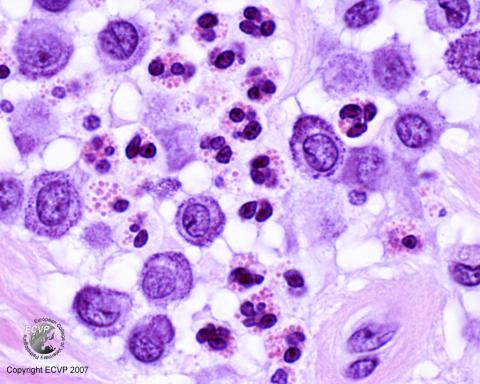
Dog skin: Mast cell tumour
Detailed information
Animal: Dog, 8 years, bull mastiff, male
Organ: Skin
History: The animal developed skin tumours on the right side of the chest and left side of the abdomen, approximately 1 cm in diameter. Histological findings: A large number of tumour cells where found growing diffusely in dermal and subcutaneous tissue. The cells were large, with moderate anisokaryosis and numerous, basophilic, cytoplasmic granules. The granules stained a metachromatic purple with the toluidine blue stain. A considerable number of eosinophilic granulocytes were found among the tumour cells.
Diagnosis: Mast cell tumour, grade 1-2
Photo by: Gjermund Gunnes