Cat: Spinal cord, lumbar segment, syringomyelia, focally extensive with associated atrophy of the gray and white matter
Animal: Cat, Ocicat, 9-years old, male neutered
Diagnosis: Spinal cord, lumbar segment, syringomyelia, focally extensive with associated atrophy of the gray and white matter
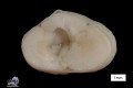
Description: Cross sections of the third lumbar segment of the spinal cord displayed an irregularly bordered liquor filled cavity, including the central left area, multifocal outreaching to the dura mater.
Comments: The cat was euthanized due to progressive ascending ataxia.
In addition to the spinal cord lesion necropsy revealed left ventricular hypertrophy of the heart with chronic congestion in lung and liver as well as amyloidosis of pancreatic islands. Histologically, a cystic cavity lacking an epithelial lining (syringomyelia) was present and accompanied by adjacent neuronal degeneration, atrophy and demyelination. Although this disorder was most likely acquired no causative inflammation, trauma or neoplasia were detectable.
Syringomyelia is a well described disorder in humans, dogs and calves and can extend over several segments of the spinal cord. Acquired forms are rare and similar to congenital forms but occur in older animals. Injury of the spinal cord, the central canal or its vascular supply due to trauma, infection or neoplasia with degeneration or cavitation of the spinal cord are proposed causes.
Picture and authored by: Kristina Dietert, Department of Veterinary Pathology, Freie Universität Berlin, Germany