Animal: Female captive bred cheetah (Acinonyx jubatus), 14 years, female
Organ: Oesophagus
History: The captive-bred cheetah lost weight and became progressively anorectic displaying a poor body condition with ruffled fur. The animal died and was submitted to necropsy.
Gross findings: The distal aspect of the thoracic oesophagus was dilated and showed increased thickness of the wall with an approx. 30 x 18 mm ulceration of the mucosa.
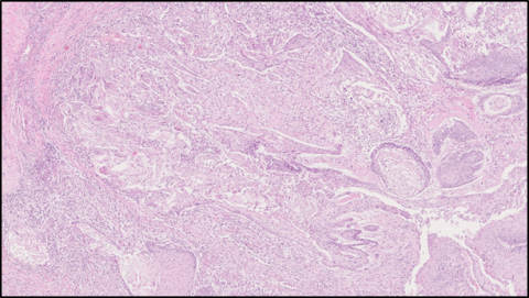
Histopathological findings: Focal irregular masses and cords of not well differentiated squamous epithelial cells, some containing intracytoplasmatic eosinophilic keratin material. Few mitosis. HE stain.
Diagnosis: Cheetah, oesophagus, squamous cell carcinoma
Necropsy performed by: Elisa Maio, Department of Pathology, Central Veterinary Research Laboratory (CVRL), Dubai, United Arab Emirates (UAE)
Photo by: Joerg Kinne, Head of Pathology Department, CVRL
Authors: Elisa Maio (CVRL), Alan Stephenson (Sheikh Butti Maktoum`s Wildlife Centre, Dubai, UAE), Joerg Kinne (CVRL)