Animal: Horse, Hanoverian thoroughbred mare, 10 years, 480 kg
Organ: hoof, left front leg
History: The horse was treated due to a pododermatitis chronica verrucosa (equine hoof canker) on the right front hoof and developed a thrombus in the left jugular vein with swelling. Subsequently, the horse showed a severe lameness on the right front leg with worsening of general condition with septic shock and final euthanasia.
Autopsy findings: Severe, widespread, transmural, acute, necro-suppurative typhlitis with thrombi, haemorrhages, vasculitis and fibrinous serositis. Thrombus within the left jugular vein, a necro-suppurative pericarditis, necrosis of renal papillae and a moderate, multifocal, erosive and ulcerative gastritis.
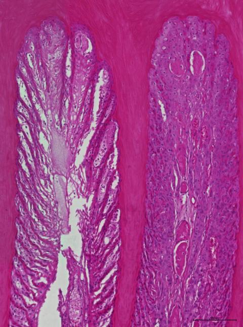
Diagnosis: Hoof, moderate, multifocal, acute necrosis of secondary epidermal lamellae with scattered neutrophil infiltration and hyaline thrombi
Necropsy performed by: Melanie Stoff
Photo by: Melanie Stoff, Vanessa Herder