Animal: Aberdeen angus, 9 months, male.
Organ: Heart
Clinical history: Multiple deaths occurred in a short time span; with short or no preceding clinical signs. When observed, clinical signs included fever, recumbency, ileus, hypersalivation. Blood exam revealed an inflammatory leukogram and increased creatin kinase.
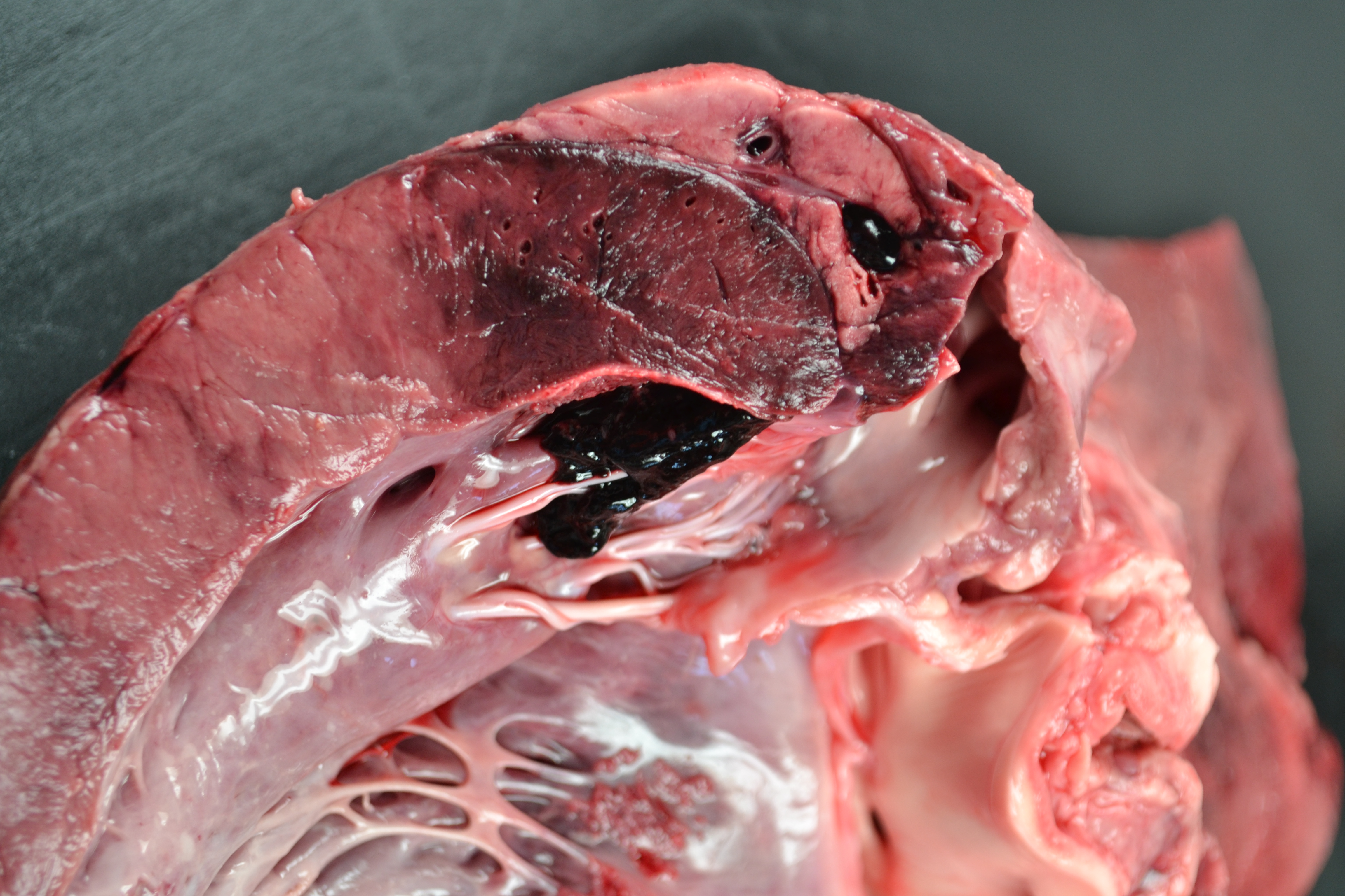
Main macroscopic findings: Marked fibrinous pericarditis; focally extensive myonecrosis and hemorrhages affecting 5-10% of the ventricular free wall (observed in the picture) and right atrium. The latter was associated with mural thrombosis. The pectoral and abdominal wall musculature showed multifocal myonecrosis, hemorrhages and emphysema (not shown).
Main histological features and diagnosis: Histologically, both the myocardial and skeletal musculature showed diffuse coagulative necrosis and expansion of the interstitia by haemorrhage, fibrin and oedema. There was occasional multifocal infiltration by neutrophils and macrophages. Rarely, low numbers of approximately 3 μm in length Gram-positive rods were scattered throughout the muscle tissue. Additionally, in the heart the epicardium was diffusely expanded by marked oedema, fibrin, hemorrhage and fewer neutrophils and macrophages.
Additional testing: Fluorescence Antibody Test (FAT) confirmed the presence of Clostridium chauvoei in impression smears of affected muscles.
Disease: Blackleg
Comment: Necrohemorrhagic myocarditis with epicarditis as well as necrohemorrhagic and emphysematous myositis with numerous intralesional Gram- positive rods is highly suggestive of clostridial myositis. Immunofluorescence confirmed the presence of Clostridium chauvoei, confirming the diagnosis of Blackleg. The heart in combination with skeletal muscle is the most common presentation (Abreu et al., 2018). The pathogenesis is thought to involve ingestion of bacterial spores, systemic distribution through bloodstream and subsequent muscle trauma would favour anaerobic environment, germination and toxin production (endogenous clostridiosis). Nonetheless, cases without direct muscle trauma as initial trigger or congenital forms have been reported (Abreu et al. 2017). Advise to the submitting veterinarian in this case included: implementation of vaccination, concomitant antibiotic treatment as well as nutritional management/moving the animals out to pasture. In this case the probable source of contamination with C. chauvoei spores was the feed/housing environment.
References
Abreu CC, Blanchard PC, Adaska JM, Moeller RB, Anderson M, Navarro MA, Diab SS, Uzal FA. Pathology of blackleg in cattle in California, 1991-2015. J Vet Diagn Invest. 2018 Nov;30(6):894-901.
Abreu CC, Edwards EE, Edwards JF, Gibbons PM, Leal de Araújo J, Rech RR, Uzal FA. Blackleg in cattle: A case report of fetal infection and a literature review. J Vet Diagn Invest. 2017 Sep;29(5):612-621. doi: 10.1177/1040638717713796. Epub 2017 Jun 9. PMID: 28599620
Contributors: Bert De Jonge, Sonja Jeckel and Bernat Marti Garcia