History: Dog, wire-haired dachshund, 1 year, female.
Diagnosis: Spinal cord (T13/L1): nephroblastoma (spinal cord tumor of young dogs) with severe neuronal compression and atrophy
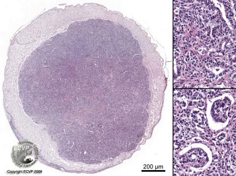
Description: Cross section of the spinal cord (T13): In the spinal cord is a well demarcated, non-encapsulated, expansile mass compressing and replacing the surrounding neuronal tissue. The neoplasm is composed of a mostly disorganized mixture of mesenchymal, epithelial and blastemal cells (see inset 1). Mesenchymal cells are haphazardly arranged in streams or partly differentiated into fibrous tissue. Epithelial cells form glandular or tubular structures and occasional tufts of cells within a lumen lined by epithelial cells (glomeruloid structures, see inset 2). Both populations blend with blastemal cells found in clumps or dispersed between the epithelial and mesenchymal components.
Comments: This young dog showed progressive paraplegia of the hind legs and was euthanized due to poor prognosis. Necropsy revealed a mass in the 13th thoracal and first lumbal segment. The histological picture in this case met the typical diagnostic criteria of a nephroblastoma (spinal cord tumor of young dogs). As in this case, tumors occur most frequently in the thoracolumbar junction. German shepherds seem to be predisposed. Because of the morphologic features and positive staining for Wilm´s tumors gene product a development of from remnants of renal tissue is assumed.
Picture by: Olivia Kershaw, Department of Veterinary Pathology, Freie Universität Berlin, Germany
Author: Olivia Kershaw, Department of Veterinary Pathology, Freie Universität Berlin, Germany