Horse: Abdominal aorta, severe, chronic, multifocal, granulomatous endarteritis
History: Horse, warmblood, female, 14 years
Diagnosis: Abdominal aorta: Severe, chronic, multifocal, granulomatous endarteritis
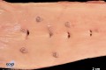
Description: Multiple, light-gray, intimal nodules of up to 1.3 cm in diameter protruded into the vascular lumen of the abdominal aorta. The lesions were located approximately 10 cm caudally to the mesenteric artery branching off the aorta.
Comments: The mare was euthanized due to a trauma to the hip. Vasculitis was an incidental finding. Clinical signs attributable to the vascular changes were absent. Histologically, the nodules appeared as multifocal chronic granulomas characterized by a central core of necrotic debris with multifocal mineralization surrounded by numerous macrophages, fewer giant cells and moderate numbers of eosinophils lymphocytes and plasma cells, peripheralized by fibrous connective tissue. Neither adult nematodes in the intestinal lumen, nor larvae within the vascular lesions were detectable. Nonetheless the granulomas were most likely caused by migrating larvae during a previous Strongylus vulgaris infection.
Picture by: Stefanie Binder, Department of Veterinary Pathology, Freie Universität Berlin, Germany
Authored by: Hannah Pischon, Department of Veterinary Pathology, Freie Universität Berlin, Germany