Animal: Cat (Felis catus), adult, female
Organ: Kidney
History: The animal was submitted to necropsy to determine the cause of death.
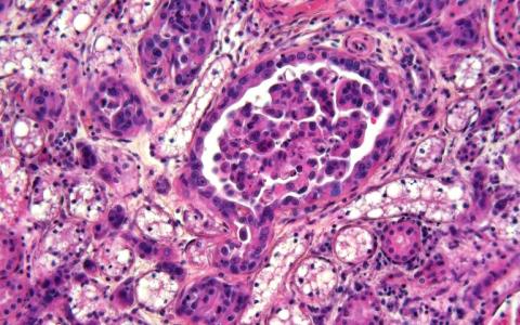
Autopsy findings: The lung showed a 10 cm in diameter, ovoid, white mass. Multiple, similar white masses with a diameter of up to 1 cm were found also in both kidneys.
Histology: Histologically, an epithelial neoplasia was found in the lung, multifocally intravascularily and randomly distributed in both kidneys. The epithelial cells were characterised by large, polygonal cells with abundant eosinophilic cytoplasm, distinct cell borders and a large nucleus with one or multiple nucleoli. There was moderate to severe anisokaryosis and -cytosis and up to 2 mitotic figures per high power field. In the lung and also in the kidney, some of the neoplastic epithelial cells displayed ciliation. Based on these findings, a pulmonary adenocarcinoma with metastasis in the kidneys is considered.
Diagnosis: Kidney, adenocarcinoma (metastatic from the pulmonary adenocarcinoma).
Necropsy and photo performed by: Maria Giovanna Cancedda, Giovanni Antonio Carboni, Ciriaco Ligios, Istituto Zooprofilattico Sperimentale della Sardegna, Sassari, Italy.