Species : Dog (Welsh Corgi), 3 months old, acute onset of vomiting, foreign body excluded. During surgery, white and hard appearance of the pylorus.
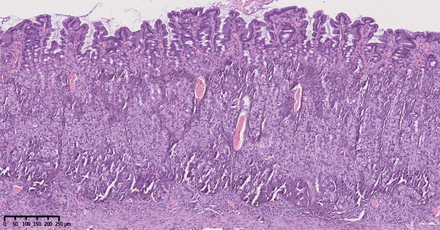
Organ : Stomach (surgical biopsies)
Description : The fundus and pylorus exhibit widespread interstitial and vascular dystrophic mineralization. It extends throughout all gastric layers, with large deposits in the medium and deep mucosa.
Morphologic diagnosis : Stomach : Multifocal severe transparietal dystrophic mineralization.
Comments : Dystrophic mineralizations of the gastric mucosa as seen here in such a young dog is most often secondary to ureamia. Juvenile nephropathy is the most likely etiology. A differential diagnosis would be rodenticide ingestion (vitamin D analogous).
Authorship (text and photos) : Charlotte Boyer (DESV-AP, Dipl ECVP) and Christelle Volmer (DMV, DESV-AP, Dipl ECVP)
Vetodiag, 6 route du Robillard, 14170 Saint Pierre en Auge, France.
cboyer@vetodiag.fr