Animal: 4-month-old goat kid.
Organ: Abomasum.
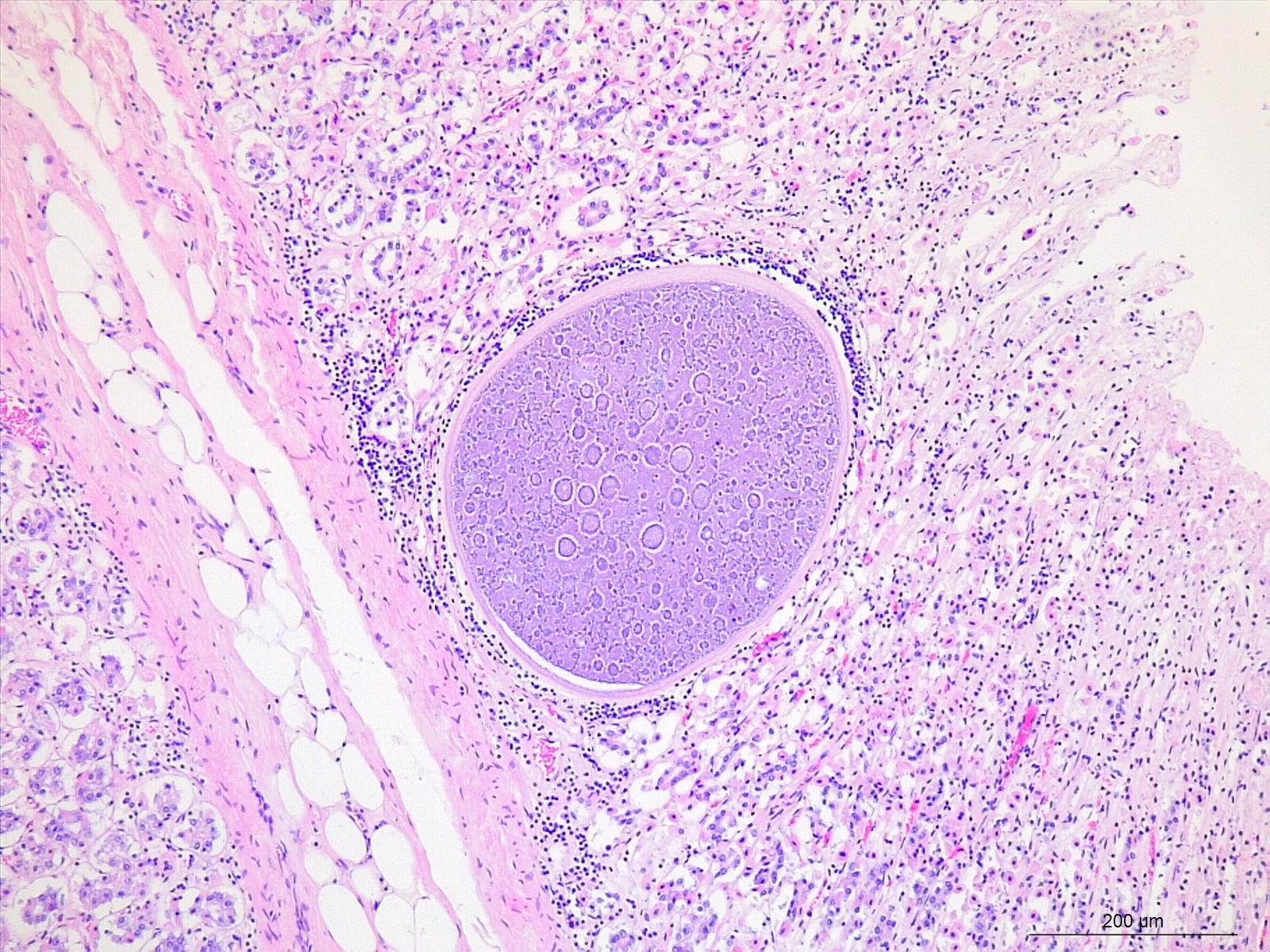
Diagnosis: Mild, multifocal, chronic, lymphoplasmacellular abomasitis with intralesional Eimeria Girluthi.
Histological features: Large schizonts (megaloschizonts) have a thick eosinophilic wall. They contain numerous of elongate merozoites. Nuclei are often arranged in circular blastophores.
Comments: Based on the histologic features and the location of the schizonts, they were classified as Eimeria gilruthi. In this case, the organism was an incidental finding during microscopic examination of the abomasum, as is commonly reported in the literature. However, this apicomplexan parasite is known to sporadically cause gastrointestinal disease in small ruminants. Clinical illness can be significant and may include diarrhea, weight loss, and even death. Animals of all ages can be affected.
Contributor: Nermin Caliskan, Animal health lab Flanders (DGZ Vlaanderen), BELGIUM.
Bibliography:
Ammar S.I., Watson A.M., Craig L.E., Cope E.R., Schaefer J.J, Mulliniks J.T., Gerhold R.W. (2019). Eimeria gilruthi–associated abomasitis in a group of ewe. Journal of Veterinary Diagnostic Investigation 31(1): 128–132.
Hermosilla C., Diakou A., Psychas V., Silva L.M.R., Taubert A. (2016). Fatal Eimeria gilruthi-induced abomasal coccidiosis: a still neglected parasitosis? Journal of Veterinary Medicine and Research 3(4): 1055.