Animal: Dog, French Bulldog, 7 years, male
Diagnosis: Cerebrum: Severe, focally extensive, chronic, granulomatous meningoencephalitis
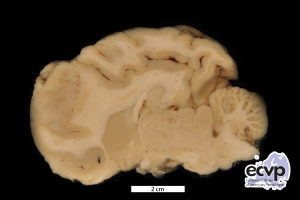
Description: A poorly demarcated, greyish mass with maximal extension of 2.1 x 1.4 x 0.8 cm was present in the frontal lobe of the telencephalon.
Comments: The dog presented clinically with ataxia of the hind limbs followed by non-responsive seizures, vomitus, apnoea and sudden death.
Necropsy revealed a greyish coloured mass in the cerebrum which presented histologically as severe granulomatous meningoencephalitis with abundant multinucleated giant cells and multifocal lymphohistiocytic perivascular cuffs. Additionally the dog had a mild, focal, chronic, lymphohistiocytic myocarditis. Special stains including Giemsa, Grocott and PAS failed to identify any pathogens.
The etiology of granulomatous meningoencephalomyelitis (GME) remains unclear. Typically, GME occurs in the cerebellum, brainstem, optic nerve or spinal cord. It is rarely seen in the cerebral cortex and white matter. Possible differentials include necrotizing meningoencephalitis (NME), necrotizing leukoencephalitis (NLE) and brain malignant histiocytosis (BMH).
Picture and authored by Anja Ostrowski, Department of Veterinary Pathology, Freie Universität Berlin, Germany