Species: Dog (Pinscher), 3 years old, cutaneous mass on the hindleg.
Organ: Skin (haired)
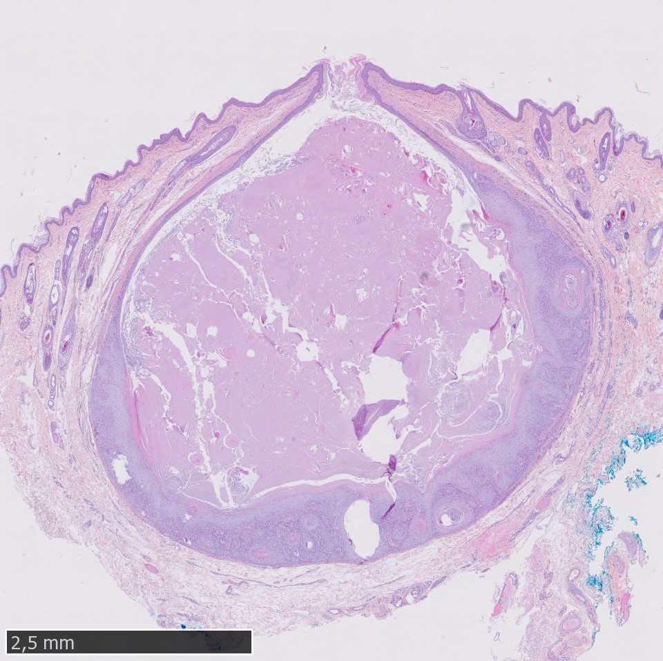
Description: A well circumscribed dermal nodule which is oriented around a central cyst filled with laminated keratin. The upper portion of the cyst is opened and connected with the epidermis. The wall of the nodule is composed of multiple small horn cysts interconnected by a reticular network of cords and trabeculae of squamous epithelium.
Morphologic diagnosis: Skin (haired) : Infundibular keratinizing acanthoma.
Comments: Infundibular keratinizing acanthoma is a benign follicular neoplasm of dogs. It accounted for 1.4 to 2.3 % of all canine skin tumors in several surveys (Stannard & Pulley, 1975 ; Goldschmidt & Shofer, 1992 ; Abramo et al.,1999). Tumors located deeper in the dermis do not have an opening of the skin surface. They are located primarily on the dorsal neck and trunk, although any part of the body can be affected.
Authorship (text and photos): Charlotte Boyer (DESV-AP, Dipl. ECVP)
Vetodiag, 6 route du Robillard, 14170 Saint Pierre en Auge, France.
cboyer@vetodiag.fr