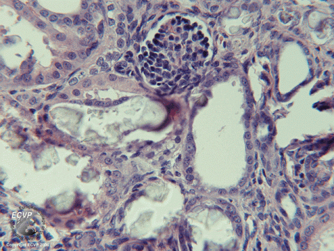
Dog Kidney: Oxalate nephropathy
Detailed information
Animal: Dog, Tibetan Spaniel, 6 weeks, female
Organ: Kidney
History: Two pups in a previous litter of three pups, had died at six-and-a-half and nine weeks of age with endstage renal lesions and massive renal tubular deposition of birefringent crystals as evident by polarisation microscopy. Both pups that exclusively had been raised by normal suckling of the healthy mother fed a normal diet, revealed signs of unthriftiness and increased thirst at five weeks of age, gradually developing to signs of disease including slight diarrhoea, inappetence, weight loss and depression (Jansen and Arnesen, 1990). This female pup was born in a subsequent litter of four pups, after remating of the identical parent animals of the previous litter (Danpure et al., 1991).
Autopsy findings: Pale, small kidneys with capsular adherence and increased tissue texture. Thin cortices containing pale, glittering material on the cut surfaces.
Diagnosis: Oxalate nephropathy. Autosomal recessive alanine:glyoxylate/serine:pyruvate aminotransferase deficiency analogous to human primary hyperoxaluria type 1.
Necropsy performed by: Johan Høgset Jansen, Norwegian School of Veterinary Science, BasAM, POB 8146 Dep., NO-0033 OSLO, Norway (jhj@veths.no).
Photo by: Johan Høgset Jansen
References
Danpure, C. J., P. R. Jennings, and J. H. Jansen, 22-2-1991: Enzymological characterization of a putative canine analogue of primary hyperoxaluria type 1. Biochim.Biophys.Acta 1096, 134-138. Jansen, J. H. and K. Arnesen, 1990: Oxalate nephropathy in a Tibetan spaniel litter. A probable case of primary hyperoxaluria. J Comp Pathol 103, 79-84.
