Animal: Dog, male, adult, Australian-shepherd crossbreed
Diagnosis: Heart, tricuspid valve: myxoma
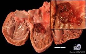
Description: An irregular, botryoid, red to brown mass measuring 3.5 x 2.5 x 2.0 cm originated from the tricuspid valve. Its surface was smooth and shiny. Histologically, the mass consisted of haphazardly arranged stellate to spindle shaped cells with elongated nuclei. The mitotic rate was low (< 1 per high power field) and the cytoplasm was slightly eosinophilic. The tumor was sustained by a fine vascular and predominantly myxoid stroma that made up to 90 % of the mass. Multifocally, mild, acute hemorrhages and areas of necrosis were present.
Comments: Myxomas are rare in the dog. These tumors are located mainly in the myocardium, particularly the left atrium. There are only few reports on other locations. Myxomas arise from stem cells that differentiate to fibroblastoid cells with synthesis of a strongly hydrated extracellular matrix. Clinically, cardiac myxomas may cause cardiac insufficiency due to incomplete closure of mitral valves with resulting, pulmonary congestion and edema, right heart dilatation and embolization.
Picture by: Lydia König, Department of Veterinary Pathology, Freie Universität Berlin, Germany
Author: Lydia König, Department of Veterinary Pathology, Freie Universität Berlin, Germany