Dog: Cerebrum, metastases of hemangiosarcoma
Dog, Labrador-Mix, 9 years.
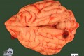
Diagnosis: Cerebrum: olfactory bulb and occipital lobe of the left hemisphere: metastases of hemangiosarcoma with focally-extensive hemorrhage and malacia
Description: The left cerebral hemisphere displayed two focal, firm, black to dark-reddishly colored masses, one of approximately 1.5 x 1.1 x 0.8 cm in the medial occipital lobe and one of approximately 0.7 x 0.6 x 0.4 cm lateral in the olfactory bulb with an uneven surface, protruding by about 0.3 cm and 0.4 cm, respectively, from the surrounding tissue. The cut surfaces were cystically structured but relatively well-demarcated.
Comments: Approximately two days prior to presentation, the dog was anorexic, depressed and experienced a singular seizure. Upon clinical presentation, symptoms included apathy, pale mucus membranes, an elevated heart rate and icteric serum. The dog died spontaneously a few hours later.
Necropsy revealed additional masses of the same consistency, color, demarcation and of similar size in the center of the Corpus callosum, in the cortex of the right kidney and, multifocally, in the caudal portions of the lung.
The primary tumor site was most likely located in the wall of right atrium at the entrance to the auricle with signs of acute rupture due to an affixed coagulum and a severe and hence fatal hemopericardium.
Histology confirmed the diagnosis of hemangiosarcoma with consecutive metastatic spread to above mentioned organs.
Hemangiosarcomas are the most common primary cardiac neoplasms in dogs, frequently involving liver, spleen and lungs with, as seen in this case, 14 % of metastases to the brain. In contrast, the occurrence of a primary intracranial hemangiosarcoma in a juvenile dog has only recently been published.
Author and image by Nancy Erickson, Department of Veterinary Pathology, Freie Universität Berlin, Germany