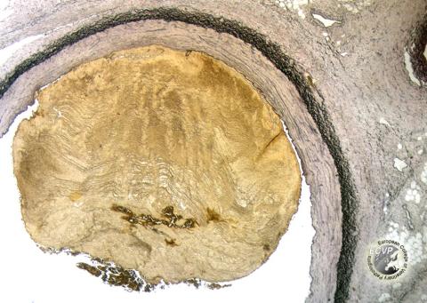
Dog, abdominal aorta: Thrombus (elastin van Gieson’s stain)
Detailed information
Diagnosis: Dog, abdominal aorta: Thrombus (elastin van Gieson’s stain).
Animal: Dog, 10 years, English Setter, female.
Organ: Aorta.
History (extract): The dog was brought to the university clinic because of sudden onset of hind leg ataxia. There was no obvious pain. The dog had been slightly depressed the last few weeks, with signs of polydipsia. There was no detectable pulse in the right femoral artery, and the distal part of the right leg was cold. In the next couple of days the signs worsened and progressed to include the left side as well. The animal was now not able to stand on its hind legs. Hypoalbuminemia and marked proteinuria were among the clinical findings. Due to a poor prognosis the dog was euthanized and submitted for autopsy.
Autopsy findings (extract): A large thrombus was found in the abdominal aorta, extending from about 5 cm orally to the pelvic inlet and into the arteria femoralis, continuing down to the knee joint on the right side and about 5 cm into the arteria iliaca externa on the left side. The thrombus was adherent to the aortic wall with degeneration of the endothelium and the lamina elastica interna. Several thrombi were also seen in the pulmonary tissue. The kidneys were swollen with an abnormally light colour. In the cortex deposits of a homogeneous, eosinophilic material were seen in most glomeruli. The material stained positively with Congo red, and was birefringent with polarised light. On electron microscopical examination, the material had a distinct fibrillar structure.
Diagnosis: Aorta: Thrombosis. It was believed that renal amyloidosis (idiopathic) was the primary lesion, causing hypercoagualbility by loss of coagulation regulatory proteins to the urine.
Necropsy performed by : Gjermund Gunnes, Norwegian School of Veterinary Science, Oslo, NorwayPhoto by: Gjermund Gunnes